Intertrochanteric Hip Fracture Case File
Eugene C. Toy, MD, Andrew J. Rosenbaum, MD, Timothy T. Roberts, MD, Joshua S. Dines, MD
CASE 5
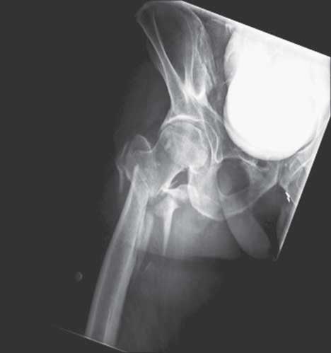
An 81-year-old white woman is brought to the emergency department (ED) with complaints of right hip pain after losing her balance while using the sink in her bathroom. Currently she is unable to bear weight on her right leg. She denies losing consciousness, feeling light-headed, or having chest discomfort or palpitations before the fall. She states her leg just “gave out.” She did not hit her head. Before her injury, she ambulated with the assistance of a cane. The patient lives alone and was unable to move herself to a phone to call for help and was not found until almost 36 hours after the fall occurred. She has no other complaints of injury. Her past medical history is significant for hypertension, hypercholesterolemia, non–insulin-dependent diabetes mellitus, hypothyroidism, osteoporosis, and a history of a proximal humerus fracture 9 months prior. On exam, the patient is lying with her right leg slightly shortened and externally rotated. She experiences significant pain with gentle passive rotation and palpation over her hip. Her skin, as well as motor and sensory function, are intact throughout her right lower extremity. Palpation of all other joints is negative for tenderness or notable deformity. Her temperature is 98ºF, blood pressure 100/60 mmHg, and heart rate is 105 beats/min. Electrocardiogram shows sinus tachycardia. An anteroposterior (AP) radiograph of the right hip is obtained (Figure 5–1).
► What is the most likely diagnosis?
► What are the treatment options available to this patient?
► What other potential medical issues must be considered based on this patient’s history?

Figure 5–1. AP radiograph of the right hip.
ANSWER TO CASE 5:
Intertrochanteric Hip Fracture
Summary: An 81-year-old woman complains of right hip pain after falling from standing and suffering a right intertrochanteric fracture.
- Most likely diagnosis: Right intertrochanteric fracture
- Treatment options: Intertrochanteric femur fractures are best treated surgically. The goal is stable internal fixation to allow early weightbearing and mobilization. Fixation implant choices include cephalomedullary hip screws, sliding hip screws, and, to a lesser extent, prosthetic replacements or external fixators. Like femoral neck fractures, nonoperative management is reserved only for patients whose medical comorbidities put them at excessive risk for surgery.
- Additional medical concerns: This patient was found collapsed, many hours after her fall. She has been without oral intake for this entire time. The patient must be evaluated for dehydration, electrolyte imbalances, nutritional deficits, pressure ulcers, potential rhabdomyolysis and associate acute kidney failure, and hemodynamic instability, as intertrochanteric fractures may be associated with a large amount of blood loss into the thigh.
- Identify and characterize intertrochanteric hip fractures with regard to their stability.
- Be familiar with the basic treatment options available for fixation of intertrochanteric fractures.
- Understand the significance of hip fractures on mortality rates.
Considerations
This patient suffered a low-energy fall from standing, with a direct impact onto her great trochanter. This represents the most common mechanism for intertrochanteric hip fractures, which are most commonly seen in elderly female patients.
On physical exam, it is important to look at the position of the affected extremity. With displaced fractures, the extremity may be shortened and externally rotated. In nondisplaced fractures, conversely, physical signs may be subtle or simply limited to pain with range of motion of the hip and an inability to bear weight. Intertrochanteric hip fractures are not commonly associated with neurovascular injury or
open fractures; however, evaluation for such characteristics is essential. In younger patients with intertrochanteric fractures secondary to high-energy mechanisms, neurovascular and associated injuries are more common. Such patients, of course, should undergo a complete and thorough advanced traumatic life support workup before orthopaedic injuries are addressed.
Several studies have shown improved patient outcomes with prompt surgical intervention for hip fractures; however, medical comorbidities must first be evaluated. In this case, the patient should be treated for dehydration and potential electrolyte imbalances and worked up for a potential rhabdomyolysis. Hip fractures are associated with increasing age (> 70 years), female sex, and low bone mass. Intertrochanteric fractures are the most common pattern, accounting for approximately 50% of all hip fractures, with femoral neck fractures being the second most frequent. Other common osteoporosis-related fractures include fractures of the vertebral bodies, distal radius, and proximal humerus. Hip fractures may also be seen in younger patients after high-energy injuries such as motor vehicle accidents and falls from height.
APPROACH TO:
Intertrochanteric Fracture
DEFINITIONS
INTERTROCHANTERIC: The region between the greater and lesser trochanters of the proximal femur. This area is extracapsular, is made up of predominantly cancellous bone, and has an abundant blood supply.
SLIDING HIP SCREW: An extramedullary device that consists of a lateral side plate and a femoral head lag screw. The lag screw is not fixed into the side plate, allowing a controlled collapse at the fracture site. This creates compression over the fracture site and may promote healing.
CEPHALOMEDULLARY NAIL: An intramedullary device that combines features of an intramedullary nail and a sliding hip screw. Cephalomedullary nails are frequently employed in unstable and reverse obliquity fracture patterns.
REVERSE OBLIQUITY: A type of unstable fracture pattern characterized by an oblique fracture line extending from the medial cortex proximally to the lateral cortex distally.
TIP-APEX DISTANCE: The sum of the distances from the tip of the lag screw to the apex of the femoral head on the AP and lateral radiographic views. The measurement is expressed in millimeters. A measurement of less than 25 mm has been shown to minimize the risk of screw cutout.
CLINICAL APPROACH
Anatomy
The femoral head is connected to the shaft by the femoral neck, which projects medially to the shaft at an angle of 120 to 130 degrees. The greater trochanter projects superiorly and laterally above the neck–shaft junction. The lesser trochanter is located at the inferior junction of the neck and shaft on the posteromedial side and is the site of insertion for the iliopsoas muscle. The 2 trochanters are connected posteriorly by a crest of thickened cancellous bone, known as the intertrochanteric crest. The piriformis and short external rotators insert along here. A strong plate of vertically oriented bone called the calcar femorale is located posteriorly between the lesser and greater trochanters. Intertrochanteric fractures may displace secondary to the pull of the musculotendinous attachments on their respective fragments: Greater trochanter fragments may be abducted and externally rotated by the gluteus medius and short external rotators, whereas the shaft will be displaced proximally, medially, and posteriorly by the adductors and hamstrings.
Mechanism of Injury
Low-energy falls from standing account for more than 90% of hip fractures in patients > 50 years of age. Fractures secondary to high-energy mechanisms commonly affect men less than 40 years of age.
Evaluation and Diagnosis
Patients most commonly present with pain in the proximal thigh and inability to ambulate after a fall. The affected extremity should be evaluated for shortening, rotation, and neurovascular integrity. Pain with log rolling or axial loading of the hip has a high association with occult fracture. Radiographic evaluation should include an AP pelvis x-ray and AP and cross-table lateral x-rays of the affected side. A traction film or an internal rotation view may assist in the visualization of subtle fractures. If a fracture is observed to extend beyond into the subtrochanteric region—defined as the region 0 to 5 cm below the lesser trochanter—full-length femur films should be acquired, as such facture patterns often require larger, longer intramedullary devices that should be planned preoperatively. When there is sufficient clinical suggestion of fracture despite normal x-ray findings, magnetic resonance imaging may be useful in establishing the diagnosis.
Classification
The Evans classification system divides intertrochanteric fractures into stable and unstable fracture patterns. In stable fracture patterns, the posteromedial cortex is intact or has minimal comminution, and thus it may still function to buttress against fracture collapse. In unstable fracture patterns, the posteromedial cortex is often too comminuted to prevent collapse of the fracture fragments, and fractures may fall into varus and retroversion without appropriate fixation. Intertrochanteric fractures with subtrochanteric extension are similarly unstable secondary to their propensity to collapse. Reverse obliquity is a special type of unstable fracture pattern in which
the fracture line extends from the proximal medial to the distal lateral intertrochanteric region. In reverse obliquity fractures, the distal fragment may displace medially secondary to both sheer forces and musculotendinous deforming forces if proper fixation is not obtained.
TREATMENT
Nonoperative Treatment
Nonoperative treatment should be considered only for patients who are nonambulatory without significant pain and those for whom surgical treatment carries an unacceptable risk of mortality. Nonsurgical management is associated with increased mortality rate, pneumonia infections, urinary tract infections, joint contractures, deep vein thrombosis, decubitus ulcers, and dementia.
Operative Treatment
Like femoral neck fractures, operative fixation is the standard of care for the vast majority of intertrochanteric fractures. Unlike nondisplaced femoral neck fractures, however, intertrochanteric fractures do not necessarily demand urgent operative intervention, because the abundant blood supply to the intertrochanteric region makes complications such as nonunion and avascular necrosis relatively rare. That stated, fracture fixation should ideally occur within the first 24 to 48 hours after fracture, as this lowers the risks associated with prolonged hospitalization and immobilization. The goal of surgery is to provide stable internal fixation to allow full weightbearing and early mobilization. The most common implants used include sliding hip screws and cephalomedullary nails.
Sliding Hip Screws: Before development of the cephalomedullary nail, the sliding hip screw was the most commonly used device for both stable and unstable fracture patterns. There are many sliding hip screw designs, but all consist of a side plate fixed to the lateral cortex of the femur and a lag screw placed into the femoral head. The lag screw should be inserted so that it is within 1 cm of subchondral bone and in a central position in the femoral head. Central placement should be assessed on both lateral and AP views. A tip-apex distance of less than 25 mm is ideal and associated with a decreased incidence of screw cutout. Recent literature has demonstrated that sliding hip screws have a higher rate of failure when used on unstable fracture patterns, especially reverse obliquity fractures. Sliding hip screws continue to have excellent outcomes in stable fracture patterns, however, with results that are at least equivalent to those of cephalomedullary nails. Theoretically, sliding hip screws allow a greater collapse and thus compression of fracture fragments as compared to cephalomedullary devices, a characteristic that is often desirable in the treatment of stable intertrochanteric fracture patterns.
Cephalomedullary Nails: This device combines features of a sliding hip screw with an intramedullary nail. Advantages include its ability to be inserted percutaneously with limited blood loss, limited fracture exposure, and decreased soft tissue damage. In addition, owing to its intramedullary location, these devices are subjected to lesser bending moments, resulting in its theoretical increased resistance to varus forces. There are several variations of the cephalomedullary nail, but all consist of an intramedullary nail and a femoral head component. The femoral head component generally consists of one or more screw or blade devices that fi x within the center of the femoral head and neck and then interlock distally with the intramedullary nail component. Insertion of the intramedullary nail may be performed through the piriformis fossa or through the greater trochanter. Piriformis nails are typically straight, as the piriformis fossa falls directly in line with the medullary canal of normal femurs; trochanteric nails have a slight lateral curve that allows them to be passed through the greater trochanter and then directed down the medullary canal. Cephalomedullary nails are best suited for unstable intertrochanteric fractures, including those with subtrochanteric extension, reverse obliquity, and comminuted posteromedial cortices. When combined with a long intramedullary component, cephalomedullary nails may also be used in the treatment of subtrochanteric fractures (ie, fractures occurring 0-5 cm distal to the lesser trochanter).
Complications
Intertrochanteric fractures, like femoral neck fractures, are associated with a high mortality rate, even when prompt operative fixation is achieved. After fracture, patients have an increased relative risk of mortality that is at least double that of their age-matched peers. Mortality rates range from 8.4% to 36% within the first year after hip fracture.
Loss of fixation is a complication seen with intertrochanteric fractures. This most commonly results from varus collapse of the proximal fragment with superior cutout of the lag screw from the femoral head. The incidence of fixation failure is reported to be as high as 20% in unstable fracture patterns. As stated, cutout may be significantly reduced when the tip-to-apex distance is < 25 mm.
Nonunion is rare, occurring in fewer than 2% of patients. It is most common in patients with unstable fracture patterns. Due to the highly vascular nature of intertrochanteric bone, however, nonunion and avascular necrosis are significantly less common after fixation in intertrochanteric fractures than in femoral neck fractures.
COMPREHENSION QUESTIONS
5.1 A patient with an intertrochanteric hip fracture undergoes open reduction and sliding hip screw application. Postoperative radiographs demonstrate that the lag screw is inferiorly and anteriorly positioned in the femoral head, with a tipapex distance of 44 mm. This patient is at greatest risk for which of the following complications?
D. Periprosthetic fracture
5.2 A 49-year-old woman with no significant past medical history is involved in a motor vehicle accident in which she sustains a reverse obliquity-type intertrochanteric femur fracture. What is the most appropriate treatment for her injury?
A. Total hip arthroplasty
B. Bipolar hemiarthroplasty
E. Cephalomedullary nail fixation
5.3 Which of the following most typically represents the patient demographic and scenario in which intertrochanteric femur fractures occur in the United States?
A. High-speed motor vehicle ejection accident in a 26-year-old man
B. Fall from standing in a 69-year-old woman
C. Fall from 3-story balcony in a 56-year-old man
D. Soccer-related internal rotation twisting injury in a 19-year-old woman
E. Twenty-year-old male pedestrian struck by a vehicle directly over the greater trochanter
ANSWERS
5.1 A. A tip-to-apex distance of less than 25 mm is associated with a decreased cutout rate of femoral head lag screws.
5.2 E. A reverse oblique intertrochanteric hip fracture is optimally treated with cephalomedullary nail fixation. Patients treated with a sliding hip screw for reverse-obliquity fractures have a much higher failure rate, nearly 56% in one study compared with 3% in the intramedullary nail group. Total hip arthroplasty, bipolar hemiarthroplasty, and external fixation are very rarely used treatment options for intertrochanteric fractures. Arthroplasty is never ideal for an otherwise healthy 49-year-old woman.
5.3 B. Although each of these mechanisms could result in an intertrochanteric femur fracture, elderly women with low-energy mechanisms most typically suffer these fractures. The average age of intertrochanteric fracture is 66 to 76 years, with published female-to-male ratios ranging from 2:1 to 8:1.
CLINICAL PEARLS
► Suspect a hip fracture in any patient presenting with a foreshortened leg, held in flexion and external rotation.
► Low-energy falls from standing account for greater than 90% of hip fractures in patients > 50 years of age.
► The stability of intertrochanteric fractures depends chiefly on the integrity of the posteromedial cortex of the proximal femur, a thick area of vertically oriented cortical bone that buttresses against fracture collapse.
|
REFERENCES
Abrahamsen B, Van Staa T, Ariely R, Olson M, Cooper C. Excess mortality following hip fracture: a
systematic epidemiological review. Osteoporosis Int. 2009;20(10):1633-1650.
Baumgaertner MR, Curtin SL, Lindskog DM, et al. The value of the tip-apex distance in predicting
failure of fixation of peritrochanteric fractures of the hip. J Bone Joint Surg Am. 1995;77:1058-1064.
Ekman EF. The role of the orthopaedic surgeon in minimizing mortality and morbidity associated with
fragility fractures. J Am Acad Orthop Surg. 2010;18:278-285.
Haidukewych GJ, Israel TA, Berry DJ. Reverse obliquity fractures of the intertrochanteric region of the
femur. J Bone Joint Surg Am. 2001;83-A(5):643-650.
Kaplan K, Miyamoto R, Levine B, Egol K, Zuckerman J. Surgical management of hip fractures: an
evidence-based review of the literature. II. Intertrochanteric fractures. J Am Acad Orthop Surg.
2008;16:665-673.
Morgan SJ. Fractures of the hip. In: Lieberman, ed: AAOS Comprehensive Orthopaedic Review. Rosemont,
IL: American Academy of Orthopaedic Surgeons; 2009:597-609.
Russell T. Intertrochanteric fractures. In: Bucholz RW, Heckman JD, eds: Rockwood and Green’s Fractures
in Adults. 7th ed. Philadelphia, PA: Lippincott Williams & Wilkins; 2010:1597-1640.


0 comments:
Post a Comment
Note: Only a member of this blog may post a comment.