Hip Osteoarthritis Case File
Eugene C. Toy, MD, Andrew J. Rosenbaum, MD, Timothy T. Roberts, MD, Joshua S. Dines, MD
CASE 40
A 65-year-old woman presents to her primary care physician with complaints of right “groin” pain that radiates to the buttock. She has noticed some mild pain in her right hip for the past 2 years, but has had severe pain over the last 4 months. She describes the pain as being “4/10” but at times can be an “8/10.” The quality of the pain is a dull ache, and she denies any weakness, numbness, or tingling in her leg below the knee; however, the pain recently has kept her from getting sleep at night. She is currently using a cane for ambulation around her community and has to rest every 4 to 5 blocks because of pain. She also notes difficulty putting on her shoes and socks and getting in and out of the car. Her symptoms are refractory to ibuprofen and acetaminophen. She denies any recent trauma, infections, or dental work. Vital signs are within normal limits. When she ambulates, she leans her body to the right side during stance phase while maintaining a level pelvis. When she is lying flat on her back, she is able to flex her right hip to 90 degrees but has severe pain past this point. She has limited abduction of her right hip compared with the left. She has 0 degrees of right hip internal rotation and 20 degrees of external rotation. When holding her left knee to her chest and extending the right hip, the right posterior thigh does not fully lie on the exam table. She has a negative straight leg raise bilaterally and is neurovascularly intact in both lower extremities.
► What is the most likely diagnosis?
► What is your next diagnostic step?
► What are the nonoperative treatment options?
► What are the surgical treatment options?
ANSWER TO CASE 40:
Hip Osteoarthritis
Summary: A 65-year-old woman presents with a chief complaint of right “groin” pain that radiates to the buttock. She is having difficulty performing activities of daily living and sleeping, and her pain is not relieved by anti-inflammatory medications. She has not had a traumatic event or recent infections. On physical exam, she has painful and limited range of motion of her hip, with a flexion contracture.
- Most likely diagnosis: Right hip osteoarthritis.
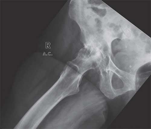
- Next diagnostic step: Anteroposterior (AP) pelvis radiograph and AP and lateral radiographs of the right hip ( Figures 40–1 and 40–2 ).
- Nonoperative treatment options: Nonsteroidal anti-inflammatory drugs (NSAIDs), physical therapy (eg, stretching and range-of-motion exercises, modalities, pool therapy), and corticosteroid injections.
- Surgical treatment option(s): Total hip replacement.
ANALYSIS
Objectives
- Understand the etiologies of arthritis.
- Be familiar with the treatment options for hip arthritis.
- Understand the indications for total hip arthroplasty.
Figure 40–1. AP pelvis radiograph demonstrating osteoarthritis in the right hip.
Figure 40–2. Frog-leg lateral radiograph of the right hip demonstrating osteoarthritic changes.
Considerations
This 65-year-old patient presents with “groin” pain, which is the typical location of pain that is coming from the hip joint. Patients often complain additionally of radiating pain to the buttock, lateral thigh, or knee. Sometimes patients who have hip arthritis will actually present with “knee pain.” This is called referred pain and is thought to be caused by irritation of the obturator nerve or, less commonly, the femoral nerve. “Hip pain” can also be due to problems in the spine and pelvis. On physical exam, pain with resisted straight leg raise and resisted hip flexion and groin pain with hip flexion and internal rotation are more commonly seen in hip disease and less commonly with low back disease. This patient also had limitations in hip range of motion, which is common with end-stage osteoarthritis. Although pain in the hip can radiate down to the knee, lumbar spine disease (eg, sciatica) will often radiate below the knee and into the foot. If the diagnosis is unclear or if the patient has both hip and lumbar spine pathology, a diagnostic injection of anesthetic and corticosteroids into the hip joint can be performed to determine whether the pain is coming primarily from the hip. If the pain is alleviated with the injection, the pain is likely coming from the hip. It is important to obtain a quality AP pelvis radiograph as well as AP and lateral radiographs of the affected hip when there is concern for arthritis ( Figures 40–1 and 40–2 ). If the patient complains of radiating pain, AP and lateral radiographs of the lumbar spine and/or weightbearing AP/lateral/Merchant x-rays of the knee may be required.
APPROACH TO:
Hip Osteoarthritis
DEFINITIONS
OSTEOARTHRITIS: Progressive, degenerative disorder of the joints caused by a gradual loss of cartilage and bone resulting in pain, stiffness, and bony overgrowth
REFERRED PAIN: Pain perceived in a body location other than the location of pathology
CLINICAL APPROACH
Etiologies
Osteoarthritis is the most common form of end-stage arthritis in the community; other types of arthritis include juvenile rheumatoid arthritis, rheumatoid arthritis, posttraumatic arthritis, inflammatory arthritis (eg, systemic lupus erythematosus), secondary arthritis from avascular necrosis, and septic arthritis. It is important to determine the etiology of the underlying arthritis, as this may affect surgical decision making and postoperative management. Radiographs are often helpful in determining the etiology of arthritis. Osteoarthritis will show evidence of asymmetric joint space narrowing, osteophyte formation, subchondral sclerosis, and subchondral cysts, whereas rheumatoid arthritis will demonstrate symmetric joint space narrowing, bony erosions, and osteopenia. Osteoarthritis of the hip may also occur secondary to previous traumatic injuries, developmental dysplasia of the hip, osteonecrosis of the hip, and femoroacetabular impingement.
Clinical Presentation
The majority of patients who present with hip pain will not require surgery. As with any painful condition, when taking a patient’s history, it is important to ask (1) location of the pain, (2) whether or not the pain radiates to another location, (3) alleviating or aggravating factors, (4) what the patient has tried to alleviate the pain, (5) quality of the pain, (6) quantity of the pain, (7) duration of the pain, (8) when the pain occurs, and (9) associated symptoms, including numbness, weakness, fevers, weight loss, involvement of other joints, and so forth.
As discussed, patients with arthritis (particularly osteoarthritis) present with groin pain that can radiate to the buttock, lateral proximal femur, thigh, or knee. Patients often describe the pain as a constant, dull ache that worsens with physical activity and is relieved, at least partially, with rest. Patients often tolerate the pain, taking anti-inflammatories or acetaminophen for many years. Often, what compels patients to finally seek treatment is restricted motion that prevents them from performing activities of daily living, including dressing and driving, or pain that interferes with their ability to sleep.
On physical exam, they often have relatively normal vital signs (unless they have an underlying medical condition) and ambulate with a coxalgic gait. A coxalgic gait is when the patient lurches his or her torso to the affected side but keeps the pelvis level. This is different from a Trendelenburg gait (a sign of hip abductor weakness), in which the patient leans his or her torso to the affected side but the pelvis is not level and is different from the antalgic gait seen with knee pathology, in which the patient limits the amount of time he or she weight bears on the affected knee during stance phase. As a result of loss of cartilage or bone, patients may have a limb length discrepancy, which may also cause an abnormal gait. With the patient supine, patients often have painful, restricted range of motion. A Thomas test, in which the patient holds his or her unaffected knee to the chest and attempts to extend the affected hip, can help the clinician to determine whether the patient also has a hip flexion contracture. Patients with hip pathology also have pain with resisted hip flexion and resisted straight leg raise. Radiographic evaluation of a painful hip includes a quality AP pelvis radiograph ( Figure 40–1 ) and an AP and lateral radiograph ( Figure 40–2 ) of the affected hip. Magnetic resonance imaging (MRI) is rarely required to make the diagnosis of arthritis, but may be beneficial in eliminating other causes of hip pain or to diagnose early cartilage loss.
TREATMENT
Total hip arthroplasty is one of the most successful procedures in medicine. Indications for total hip replacement in general terms are debilitating, end-stage arthritis of the hip that limits activities of daily living. Orthopaedic surgeons generally evaluate pain, success (or lack of success) of nonoperative interventions, baseline function, mobility, and radiographic evidence of joint space damage in their decision to recommend surgery. In addition, patients must have failed nonoperative treatment interventions, including physical therapy (eg, stretching and range-of-motion exercises, modalities, pool therapy), nonsteroidal anti-inflammatories, and intraarticular corticosteroid injections.
A variety of implants and bearing surfaces are currently available. The most common current bearing options include metal on metal, metal on polyethylene, ceramic on ceramic, ceramic on-polyethylene, and Oxinium on polyethylene. With each of the previously mentioned types of bearings, the femoral head is former and the acetabular liner is latter. There are advantages and disadvantages to each type of bearing surface. In older adults, the most commonly used bearing surface is metal on polyethylene. In addition, both the acetabular and femoral components can be cementless or cemented, depending on the patient’s age and bone quality and surgeon preference.
COMPREHENSION QUESTIONS
40.1 A 58-year-old golfer presents to your office with complaints of progressive right groin pain, 2 years in duration. Lately he has been having difficulty walking between holes on the golf course. On examination, he is overweight, exhibits an antalgic gait, and has markedly limited range of hip motion. Which of the following choices will provide the most information to the orthopaedic surgeon regarding the presence of degenerative joint disease?
A. AP pelvis plain x-rayB. CT scan of the pelvisC. AP pelvis, AP, and lateral plain radiographs of the affected hipD. AP and lateral comparison plain x-rays of the unaffected hipE. MRI of the pelvis
40.2 A 68-year-old male active tennis player presents to your practice with a 4-year history of left groin pain. His pain has been worsening over the last year, and he is now unable to participate in the local singles league. He has moderate relief with NSAIDs and acetaminophen in the past, but states these are no longer fully effective. An orthopaedic spine surgeon has evaluated him in the recent past for degenerative disc disease but only treated him with NSAIDs and prednisone for what he recalls as a “slipped disc.” On exam, the patient has a limited range of hip motion and pain with internal rotation. Straight leg raise above 70 degrees causes pain to radiate down his leg. X-rays show mild degenerative changes in his bilateral hips. What is the next best step in management of this patient?
A. Left total hip arthroplastyB. Left hip hemiarthroplastyC. Immediate referral back to his spine surgeon for further workupD. Intraarticular corticosteroid injectionE. Continue NSAIDs and acetaminophen, recommend physical therapy, and add a low-dose narcotic for daily pains
40.3 A 63-year-old obese woman is referred to your office for evaluation for arthritis, diagnosed on x-rays ordered by her primary care physician. The patient complains that she gets left hip pain when power-walking with her dog. She states she has an aunt who had relief of chronic pain from a hip replacement last year and wants you to perform the same “miracle operation.” A careful history, physical exam, and appropriate imaging confirm the diagnosis of left hip osteoarthritis. Which of the following is not an appropriate first-line treatment?
A. Activity modificationB. Weight lossC. Physical therapy for quadriceps strengtheningD. Intraarticular steroid injectionE. Total hip arthroplasty
ANSWERS
40.1 C. AP pelvis, AP hip, and frog-leg lateral radiographs are the standard films to obtain of the hip when evaluating a patient for degenerative joint disease. CT scans and MRI are typically unnecessary in the setting of osteoarthritis.
40.2 D. It is unclear by this question whether his symptoms are from osteoarthritis or spinal pathology. In these situations, intraarticular corticosteroid injections may play a diagnostic role in addition to being therapeutic. The simultaneous injection of lidocaine or similar local anesthetics should provide near-immediate relief of symptoms if his pain is arthritic in nature. Although referral and/or consultation with a spine surgeon is appropriate, this patient may well have symptomatic osteoarthritis of the hip and should not be simply sent to see someone else. Although NSAIDs, acetaminophen, and physical therapy play an important role in his therapy, they do not necessarily provide the diagnostic evidence needed to determine the source of his pain; secondary, chronic narcotics should almost never be used in the treatment of degenerative joint disease. Of course, total hip arthroplasty or hemiarthroplasty should not be performed until the surgeon is certain of the underlying diagnosis and has exhausted nonoperative treatments when effective.
40.3 E. Activity modification, physical therapy (especially that directed at quadriceps strengthening), weight loss, and intraarticular steroids are all first-line nonoperative treatments for osteoarthritis. Total hip arthroplasty is reserved for patients who have failed conservative measures and whose arthritis is adversely affecting their ability to accomplish activities of daily living.
CLINICAL PEARLS
|
► The most common cause of end-stage hip arthritis in the United States is osteoarthritis. ► Most patients with intraarticular hip pathology present with groin pain that may or may not radiate to other locations around the hip joint or the knee. ► If it is unclear whether or not a patient is having hip, back, or pelvis pain, a diagnostic intraarticular injection of anesthetic and corticosteroids may help elicit the source of the underlying pain. ► Standard radiographic imaging of patient with hip arthritis includes an AP pelvis radiograph and an AP and lateral radiograph of the affected hip. ► First-line therapies for degenerative disease of the hip are almost always nonoperative in nature. They include interventions such as physical therapy (eg, stretching and range-of-motion exercises, modalities, pool therapy), nonsteroidal anti-inflammatories, and intraarticular corticosteroid injections. |
REFERENCES
Hochberg MC, et al. Guidelines for the medical management of osteoarthritis. Part I. Osteoarthritis of the hip. American College of Rheumatology. Arthritis Rheum . 1995;38:1535-1540.
Huo MH, et al. What’s new in total hip arthroplasty. J Bone Joint Surg Am . 2011;92:2959-2972.
Pluot E, et al. Hip arthroplasty. Part 1: prosthesis terminology and classification. Clin Radiol. 2009;64: 954-960.


0 comments:
Post a Comment
Note: Only a member of this blog may post a comment.