Achilles Tendon Rupture Case File
Eugene C. Toy, MD, Andrew J. Rosenbaum, MD, Timothy T. Roberts, MD, Joshua S. Dines, MD
CASE 34
A 35-year-old man is brought to the emergency department (ED) by his basketball teammates for evaluation of his right leg. The patient was playing a pick-up game at the local gym earlier in the day, at which time he planted his right foot for a jump shot and suddenly felt as though someone kicked him in the back of his leg, although when he looked back no one was close by. The patient reports feeling a “snap” in his calf, experiencing intense pain, and falling to the ground. In the ED, his vital signs are within normal limits. Examination of his left lower extremity reveals a palpable, tender “bulging” mass over his calf and a soft depression on the posterior aspect of his heel. There is no bony tenderness of the left lower extremity. On passive range of motion, there is increased dorsiflexion in the left foot compared with the right foot. The patient cannot actively plantarflex his left foot.
► What is the most likely diagnosis?
► What is the next step in workup?
► What is the best treatment?
ANSWER TO CASE 34:
Achilles Tendon Rupture
Summary: A 35-year-old man presents with left lower extremity pain after hearing a “snap” in the back of his leg while planting his foot to take a shot during a basketball game. He states that it felt like someone kicked him. Immediately afterward he noticed significant posterior heel swelling. He is unable to actively plantarflex his foot. On exam, you note increased passive range of dorsiflexion on his left side when compared with his right as well as a palpable defect in his Achilles tendon.
- Most likely diagnosis: Achilles tendon rupture.
- Next step in workup: Pain control, plain radiographs and magnetic resonance imaging (MRI) of the left distal tibia and fibula and ankle if the diagnosis is unclear. Ultrasound is an effective alternative modality for diagnosing an Achilles tendon rupture if MRI is unavailable or contraindicated.
- Best treatment: Primary surgical repair.
- Understand how to clinically diagnose an acute Achilles tendon rupture using history and specific physical exam maneuvers.
- Be familiar with treatment options, both nonoperative and operative, for managing Achilles tendon ruptures.
- Recognize the indications for various treatment options and understand their respective advantages and disadvantages.
Considerations
Initial treatment for this 35-year-old man with a presumed Achilles tendon rupture should focus on pain relief and placing the affected leg in a splint that ensures immobilization in a plantarflexed position. The goal of this initial stabilization is to restore the natural length of the musculotendinous unit disrupted by the injury. The primary goal of treatment is to alleviate pain and restore function. There is debate in the orthopaedic community regarding the best treatment modality for acute Achilles tendon ruptures. Nonoperative treatment includes a period of 6 to 8 weeks of splint or cast immobilization. Operative surgical treatments include open tendon repair and minimally invasive suturing techniques to restore the length and integrity of the torn tendon.
APPROACH TO:
Achilles Tendon Rupture
DEFINITIONS
THOMPSON TEST: With the patient in the prone position, the examiner, squeezes the posterior calf musculature ( Figure 34–1 ). A positive Thompson test occurs when there is an absence of plantarflexion in the affected extremity. The disrupted musculotendinous unit is no longer able to plantarflex the foot on the affected side. This is compared with the unaffected side.
ECCENTRIC CONTRACTION: A muscular contraction during which muscle fibers actually elongate while firing due to overwhelming antagonistic forces. This occurs because the forces that the muscle is trying to overcome are greater than the force generated by the muscle body. Conversely, concentric contractions occur when the muscle body shortens with contraction. In concentric contractions, the force generated by the muscle is greater than the antagonistic forces applied to it.

Figure 34–1. Thompson test, in which compression of the gastrocnemius-soleus complex normally
produces plantarflexion of the foot (1). If the tendon is completely ruptured, this will not occur (2).
(Reproduced, with permission, from Knoop KJ, Stack LB, Storrow AB, et al. Atlas of Emergency Medicine. 3rd ed. New York, NY: McGraw-Hill; 2009:Fig. 11-77.)
CLINICAL APPROACH
Etiologies
Diagnosis of Achilles tendon ruptures are increasing in incidence as more “weekend warrior” athletes are pushing the limits of their typically sedentary and deconditioned bodies. Additionally, the medical community is becoming more aware of and skilled at diagnosing the injury. This injury is commonly observed in men in their fourth through sixth decades of life.
Pathoanatomy
The gastrocnemius and soleus muscle bodies are contained within the superficial posterior compartment of the leg. These muscle bodies converge to form a common Achilles tendon, which inserts on the posterior tuberosity of the calcaneus. This musculotendinous unit acts to plantarflex the foot. The Achilles tendon most commonly ruptures when the gastrocnemius-soleus complex undergoes eccentric loading with the ankle dorsiflexed and the knee extended. Patients are usually able to describe the incident in which they were pushing off or landing on a plantarflexed foot. Most Achilles tendon injuries occur in a hypovascular watershed area located approximately 2 to 6 cm proximal to its insertion. Repetitive microtrauma to this vulnerable hypovascular region is thought to predispose it to rupture.
Physical Exam
Along with the clinical history, the physical exam is key to diagnosing an Achilles tendon rupture. Despite increasing awareness in the medical community, approximately 25% of Achilles tendon ruptures are initially misdiagnosed as ankle sprains. Visual inspection of the affected extremities will likely reveal significant soft tissue swelling in the heel region. A palpable gap, erythema, and/or a bulging muscle mass may be noted after an acute rupture. The Thompson test ( Figure 34–1 ) will aid the clinician in establishing the diagnosis of an Achilles tendon rupture. Although plantarflexion is frequently compromised, be aware that the patient may still exhibit active plantarflexion due to the functions of the unaffected toe flexor and posterior tibialis muscles.
Imaging
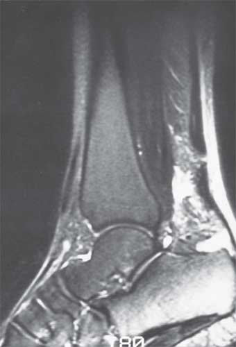
The diagnosis of an Achilles tendon rupture is primarily a clinical diagnosis. Imaging studies are generally reserved for situations in which the physical exam is inconclusive. Plain radiographs of the lower leg have limited use in diagnosing soft tissues injuries but can adequately rule out any bony injuries and sometimes demonstrate soft tissue swelling. Ultrasound is another imaging modality that can be useful. Although inexpensive, its utility is greatly operator dependent. MRI is the gold standard imaging modality but is not necessary in a clinically obvious diagnosis of Achilles tendon rupture ( Figure 34–2 ). It may, however, be useful in discerning subtleties of partial or chronic Achilles tendon injuries. Of note, the American Academy of Orthopaedic Surgeons Clinical Practice Guideline Summary for acute Achilles tendon rupture states that “We are unable to recommend for or against the routine use of MRI, ultrasonography, and radiography to confirm the diagnosis of acute Achilles tendon rupture.” In other words, it remains a clinical diagnosis.

Figure 34–2. Sagittal T2-weighted MRI image of an Achilles tendon rupture. Note the hyperintensity
surrounding its calcaneal insertion that is indicative of edema and an absence of tendinous attachment.
(Reproduced, with permission, from Skinner HB. Current Diagnosis & Treatment in Orthopedics.
4th ed. New York, NY: McGraw-Hill; 2006:Fig. 9-58.)
TREATMENT
The treatment for an Achilles tendon rupture is controversial and should be individualized to each patient. The primary goal is to reestablish the normal anatomy of the muscle and tendon, in turn restoring function to the musculotendinous unit. If the tendon is left to heal in either extreme of dorsiflexion or plantarflexion, the mechanical functioning of the gastrocnemius-soleus complex with the tendon could be compromised.
Nonoperative treatment is reserved for older, sedentary individuals with multiple medical comorbidities that may compromise wound healing. It focuses on immobilizing the affected leg in a gravity equinus position that maximizes apposition of the 2 ends of the ruptured tendon. Posterior splints, removable boots and short leg casts for 6 to 8 weeks can all stabilize the foot in this manner. Gradual range of motion and resistance exercises begin at 8 to 10 weeks. Such management lacks the risks of surgery, which include infection, wound breakdown, and nerve injury. However, the limitations of conservative management include suboptimal tendon length after healing and diminished muscle function. Patients must be warned that they will likely have residual weakness and that it can take up to 1 year before maximal plantarflexion is achieved. Furthermore, some studies have shown an increased re-rupture rate with nonoperative management.
Common indications for operative treatment include acute ruptures in young, healthy, active individuals, and following re-rupture of previously immobilizationtreated Achilles ruptures. It can be accomplished via open and percutaneous techniques. The surgical approach is on the medial side of the Achilles tendon sheath. The frayed edges are debrided, and the foot is positioned in equinus. Two heavy nonabsorbable sutures are then woven through 3 to 4 cm of each tendon edge, using a Bunnell or Kessler stitch technique. The repair can be augmented with lighter, absorbable sutures or with the plantaris tendon if it is present. Although surgical repair decreases re-rupture rate and increases maximal plantarflexion strength, it does have risks and complications, including infection, skin sloughing and necrosis, and sural nerve damage.
COMPREHENSION QUESTIONS
34.1 A 28-year-old man with an acute Achilles tendon rupture is deciding between operative and nonoperative treatment for his condition. Which of the following statements is true regarding operative treatment?
B. Lower incidence of tendon re-rupture
C. Decreased muscle strength
D. Increased sensation over the skin of the posterior calf
34.2 A 45-year-old man presents with severe ankle pain and an inability to bear weight after tripping into a sand trap while golfing. Which initial diagnostic test is most appropriate?
34.3 Achilles tendon rupture typically occurs during which type of muscle contraction?
A. Eccentric muscle contraction
B. Isometric muscle contraction
C. Concentric muscle contraction
D. Rhythmic muscle contraction
ANSWERS
34.1 B. Surgical repair is associated with a significantly reduced risk of re-rupture when compared with nonsurgical treatment.
34.2 B. In a patient with ankle pain and an inability to bear weight, potential fracture must first be ruled out. Although MRI is the gold standard for diagnosing Achilles tendon ruptures, MRI, and similarly ultrasound, should be considered only after other potential injuries are excluded. Bone scans are effective at diagnosing occult bony abnormalities but rarely play a role in diagnosing acute injuries about the ankle.
34.3 A. The Achilles tendon most commonly ruptures when the gastrocnemiussoleus complex is undergoing eccentric loading with the ankle dorsiflexed and the knee extended.
CLINICAL PEARLS
|
► Approximately 25% of Achilles tendon ruptures are initially misdiagnosed as ankle sprains.
► Signs and symptoms of an acute rupture include pain, swelling, and ecchymosis around the heel, and a palpable gap in the tendon. Patients classically recall a “popping” sensation during injury or describe the false sensation of being struck on the heel with a blunt object.
► The Thompson test is an effective physical exam maneuver used to diagnose Achilles ruptures.
► While Achilles tendon rupture is a predominantly clinical diagnosis, imaging may be necessary in the setting of chronic symptomatology, potential concomitant injuries, or unclear exam findings.
|
REFERENCES
Azar FM. Traumatic disorders. In: Canale ST, Beaty JH, eds. Campbell’s Operative Orthopaedics. 11th ed.
Philadelphia: Mosby Elsevier; 2008:2737-2788.
Casillas MM, Mann RA. Tendon disorders of the foot and ankle. In: Chapman MW, Szabo RM, Marder
R, et al, eds. Chapman’s Orthopaedic Surgery. 3rd ed. Philadelphia: Lippincott Williams & Wilkins;
2001:3123-3136.
Chiodo CP, et al. AAOS Clinical practice guideline summary: diagnosis and treatment of acute Achilles
tendon rupture. J Am Acad Orthop Surg. 2010;18:503-510.
Chiodo CP, Wilson MG. Current concepts review: acute ruptures of the Achilles tendon. Foot Ankle Int.
2006;27:305-313.
Nilsson-Helander K, et al. Acute Achilles tendon rupture: a randomized, controlled study comparing
surgical and nonsurgical treatments using validated outcome measures. Am J Sports Med. 2010;38:
2186-2193.


0 comments:
Post a Comment
Note: Only a member of this blog may post a comment.