Slipped Capital Femoral Epiphysis (SCFE) Case File
Eugene C. Toy, MD, Andrew J. Rosenbaum, MD, Timothy T. Roberts, MD, Joshua S. Dines, MD
CASE 17
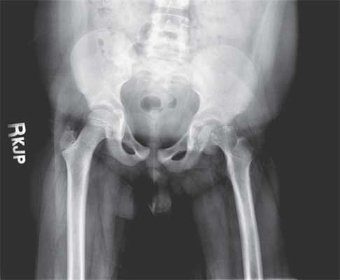
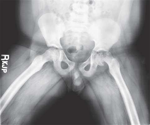
A 13-year-old African American male patient presents to the emergency department with pain in his left knee after falling on his side during a soccer game. He is unable to ambulate and is in significant discomfort. His mother states that he has been experiencing several weeks of pain in his left knee before his fall, but x-rays and repeated exams of his knee at his primary care physician’s office had failed to demonstrate any pathology. The patient has no known past medical history. He denies fevers, chills, and recent illness, and he recalls no history of traumatic injury to his left lower extremity. On examination, the child is obese and is comfortable after administration of appropriate analgesia. His left lower extremity is held in slight external rotation. His knee is without effusion and has a full range of painless motion. Motion at the hip, however, is painful, especially with passive internal/external rotation. He is neurovascularly intact throughout his bilateral extremities. Exam of the right lower extremity is unremarkable. Laboratory studies are significant for a thyroid-stimulating hormone (TSH) level of 7.3 mIU/mL (normal 0.6-5.5 mIU/mL). An anteroposterior (AP) pelvis and bilateral frog-leg lateral views are shown in Figures 17–1 and 17–2 , respectively.
► What is the most likely diagnosis?
► What aspects of this patient’s history put him at risk for this injury?
► What is the next step in the management of this patient?
Figure 17–1. AP radiograph of the pelvis.
Figure 17–2. Frog-leg lateral radiograph of the pelvis.
ANSWER TO CASE 17:
Slipped Capital Femoral Epiphysis (SCFE)
Summary: A 13-year-old obese, African American male presents with several weeks of left lower extremity pain, exacerbated by a recent fall. Exam demonstrates left hip pathology that is referred to the knee. Additionally, the patient has previously undiagnosed hypothyroidism.
- Most likely diagnosis: Left slipped capital femoral epiphysis.
- Historical risk factors: Obesity, male, African American, adolescence, with concomitant endocrinopathy (hypothyroidism).
- Next appropriate step: Percutaneous screw fixation.
ANALYSIS
Objectives
- Recognize the presentation of SCFE.
- Understand the workup for SCFE and patient population.
- Be familiar with the treatment for SCFE and its potential long-term complications.
Considerations
This is an overweight, African American male adolescent with a history of pain in his left knee. Up to 46% of patients with SCFE will present initially with complaints of distal thigh or knee pain. It is essential to recognize that hip pathology can often be referred to the distal thigh or knee. Complaints of knee pain should warrant complete and thorough physical exam, and typically radiographic evaluation, of the ipsilateral hip. Patients with SCFE typically present with an externally rotated, subtly foreshortened lower extremity, with a markedly decrease range of painful internal rotation.
Hip x-rays should include, at a minimum, an AP view of the pelvis as well as lateral views of each femoral head and neck, typically achieved through a frog-leg lateral view. Although this patient’s SCFE is clearly visible on both AP and lateral films, early SCFE lesions are often subtle and are generally apparent on lateral views before they are obvious on the AP.
Patients with newly diagnosed SCFE lesions should be immediately made nonweightbearing on the effected extremity. Complete workup for SCFE secondary to underlying medical conditions should be performed, including initial laboratory testing for TSH, a complete metabolic panel, and a complete blood count. Patients undergoing potential operative fixation should always receive blood type and screening and coagulation (prothrombin time/partial thromboplastin time) studies.
APPROACH TO:
Slipped Capital Femoral Epiphysis (SCFE)
DEFINITIONS
OSTEONECROSIS: The cellular death of bone, typically resulting from a prolonged disruption of blood supply.
STABLE SCFE: Defined simply by the patient’s ability to ambulate, even with crutch assistance. Less than 10% of patients with a stable SCFE develop osteonecrosis.
UNSTABLE SCFE: Defined as SCFE in patients who are unable to ambulate. These patients have a high incidence of osteonecrosis, upward of 50%.
CLINICAL APPROACH
SCFE (often pronounced “skiffy”) is a disorder of adolescence in which a fracture— or, technically, a disruption—occurs through the growth plate of the femoral head. The epiphysis, or region of developing bone above the growth plate (physis), is therefore mobile and tends to “slip” from the neck of the femur under the repetitive loading of body weight. The “slipping” process is actually a misnomer, however, as it is not technically the epiphysis that slips from the femur, but rather the femur that displaces from the anatomically stable epiphysis.
Epidemiology
SCFE typically affects children between 10 and 17 years of age, occurring at an average of 13.4 years for boys and 12.2 years for girls. Its prevalence in the United States is 10 in 100,000 people annually. SCFE has a slight male-to-female predominance of 3:2 and occurs at increased incidences of 2.2 in patients of African ancestry, 4.5 in those of Pacific Islander ancestry, and 0.1 for North African and Indian subcontinental ancestry, versus 1.0 for white controls. Racial differences are closely related to average adolescent body weights.
Pathogenesis
As stated, SCFE is a failure of the physis, with separation of the epiphysis from the proximal femoral metaphysis. Biomechanical factors such as obesity, femoral retroversion (increased posterior angulation of the femoral neck), and increased physeal obliquity (an increasingly angulated growth plate that is vulnerable to shearing forces when body weight is loaded) all contribute to physeal weakening. In younger children, the physis is protected by a perichondral ring that resists shearing forces. This protective ring weakens in adolescence, however, and increases the risk of SCFE. SCFE occurs during puberty, when rapid cellular expansion is occurring at the physeal zone of hypertrophy. Failure is thought to occur at this slightly weakened zone of rapid, immature expansion. Finally, hormonal and endocrine changes are associated with SCFE, but the mechanisms by which they contribute to the disease process are not fully understood. There are data to show that hypothyroidism, growth hormone supplementation, and hypogonadism increase one’s risk of SCFE.
Radiology
The direction of a typical “slip” causes the femur to fall into varus, extension, and external rotation. Typically, the epiphysis tends to move posteriorly first, a translation that is most apparent on frog-leg lateral views. With the femurs externally rotated, abducted, and flexed, an unobstructed lateral view of the femoral neck shows early posterior displacement of the epiphysis. For this reason, frog-leg lateral images are considered most sensitive for the diagnosis.
Several radiographic measurements can be made to diagnose and grade SCFE. The Klein line, or a line drawn parallel to the superior femoral neck, should intersect the epiphysis in normal individuals. In patients with advanced slips, however, the Klein line contacts the edge of, or is superior to, the migrating epiphysis. The metaphyseal blanch sign of Steel is a blurring of the proximal femoral metaphysis that may be visualized on an AP pelvis film. This is caused by overlapping of the normal metaphysis with the posteriorly displaced epiphysis.
CLASSIFICATIONS
Although several classification schemes exist, the most practical and prognostically relevant classification divides the disease into stable and unstable SCFE. SCFE stability is defined simply by whether or not the patient is able to tolerate weightbearing on the affected extremity. Stability includes those who are able to partially weight bear with crutches. Patients with unstable SCFE are so uncomfortable with movement of the hip that they refuse to ambulate. Up to 50% of patients with unstable SCFE have an incidence of osteonecrosis of the femoral head.
TREATMENT
Intervention should occur as soon as the diagnosis is made. For patients with mild or moderate stable disease, in situ fixation is the method of choice. Attempts to forcefully reduce the deformity are not recommended; however, sometimes the slip will spontaneously reduce or improve when the patient is positioned for surgery. Regardless, the goal or treatment is to stabilize the slipped epiphysis, as remodeling often occurs and patients can tolerate a certain degree of residual external rotation. Single-screw percutaneous fixation is the most common mode of treatment, although double-screw fixation techniques are sometimes performed. For singlescrew fixation, the goal is to place the screw through the middle of the epiphysis and perpendicular to the growth plate. Patients with stable slips are typically allowed to bear weight after fixation. Bilateral fixation may be indicated in patients with underlying endocrinopathies, even if the contralateral hip is asymptomatic and without radiographic evidence of the disease. Although controversial, some authors also advocate for the prophylactic pinning of the contralateral hip in children less than 10 years of age with unilateral SCFE or in those with open triradiate cartilage. The impetus behind prophylactic fixation of the unaffected hip stems from the elevated rates at which young children and patients with endocrinopathies develop bilateral disease.
In patients with unstable SCFE, there is significant controversy over whether reduction manipulations should be employed versus in situ fixation, whether capsulotomy or arthrocentesis should be performed versus no joint decompression, and whether single- versus multiple-screw fixation techniques should be used. Most surgeons advocate relatively urgent treatment in these patients, as greater than 24 hours between acute injury and fixation may be associated with increased risk of osteonecrosis. A common treatment regimen for unstable SCFE involves single-screw fixation after joint aspiration to relieve intracapsular pressure and promote vascular perfusion. These patients are generally made nonweightbearing with crutches for 6 to 8 weeks postoperatively.
Complications
Osteonecrosis is a severe, debilitating complication, for which risk is increased with unstable SCFE, delayed surgical fixation of acute unstable SCFE, attempted reduction manipulations, and improper placement of pins, specifically in the posteriorsuperior femoral neck, leading to disruption of vasculature. Osteonecrosis is initially managed with nonweightbearing, nonsteroidal anti-inflammatory drugs, and gentle range-of-motion exercises. When severe, however, reconstructive intervention may be necessary. Slip progression after initial fixation is another potential complication that, fortunately, occurs in only 1% to 2% of patients after single-screw fixation. Although double-screw fixation may theoretically reduce this complication, elevated potential risks of osteonecrosis with multiple-screw fixation favors singlescrew techniques for most surgeons.
COMPREHENSION QUESTIONS
17.1 A 13-year-old boy is referred to your office with a right-sided SCFE after workup over several weeks by his pediatrician for insidious right knee pain. On exam, he has obligatory external rotation with hip flexion and is refusing to bear weight. His TSH is 8.5 mIU/mL. Which of the following is the most appropriate treatment for this patient?
A. Percutaneous pinning of the right hipB. Open reduction and capsulotomy of the right hip with plate and screw fixationC. Percutaneous pinning of the left hipD. Percutaneous pinning of bilateral hipsE. Administration of levothyroxine with follow-up TSH levels in outpatient setting and bed rest until resolution of pain
17.2 Through which of the following physeal zones does the disruption in SCFE typically occur?
A. Reserve zoneB. Proliferative zoneC. Hypertrophic zoneD. Zone of provisional calcification
17.3 An 8-year-old obese girl of Pacific Island ancestry is referred to your office for a right-sided unstable SCFE lesion after a fall while playing kick-ball. She had no previous pain in this extremity. On examination, she has an externally rotated right lower extremity that is severely painful with passive range of motion. Frog-leg lateral x-rays show a displaced right SCFE lesion with a normal-appearing left femoral head. Which of the following factors is indication to prophylactically fix this patient’s left hip?
A. ObesityB. AgeC. Unstable nature of the right-sided SCFED. Acute, traumatic right-sided SCFE without preexisting symptomsE. Pacific Island ancestry
ANSWERS
17.1 D. Bilateral percutaneous screw fixation is appropriate in this patient due to the patient’s history of undiagnosed hypothyroidism. His right hip, most urgently, requires fixation, but the presence of endocrinopathy is a generally accepted indication for additional fixation of the unaffected contralateral side. Management of hypothyroidism is important in this patient, but bed rest is generally an unsuitable treatment for SCFE.
17.2 C. SCFE classically occurs through the zone of hypertrophy, a region of rapid cellular expansion during the adolescent growth spurt that is especially vulnerable to shearing injury.
17.3 B. Indications to prophylactically fix the contralateral hip in a patient with a unilateral SCFE lesion are limited to patients with obvious endocrinopathies and those younger than 10 years of age or with open triradiate cartilage. This patient’s age is reason enough to strongly consider preemptively pinning the left hip. Her obesity and ethnicity are risk factors for SCFE; however, they are not indications for prophylactic fixation. The unstable and acute nature of her SCFE lesions does not necessarily correlate with a need for prophylactic intervention.
CLINICAL PEARLS
|
► Remember that hip pathology can often be referred to the distal thigh or knee and thus a complete exam of both joints is essential. Almost 50% of SCFE patients present with distal thigh or knee pain. ► Percutaneous single-screw in situ fixation is the treatment of choice for SCFE. ► Prophylactic fixation of the contralateral hip, regardless of whether or not it is symptomatic, is generally advocated in patients with SCFE secondary to endocrinopathies or in SCFE patients younger than 10 years at presentation. Admittedly, this is controversial. |
REFERENCES
Aronsson DD, Loder RT, Breur GJ, Weinstein SL. Slipped capital femoral epiphysis: current concepts. J Am Acad Orthop Surg. 2006;14:666-679. Flynn, JM, ed. Hip, pelvis, and femur disorders: pediatrics. In: Orthopaedic Knowledge Update: Ten. Rosemont, IL: American Academy of Orthopaedic Surgeons; 2011:739-752.


0 comments:
Post a Comment
Note: Only a member of this blog may post a comment.