Ulnar Collateral Ligament Injury of the Elbow Case File
Eugene C. Toy, MD, Andrew J. Rosenbaum, MD, Timothy T. Roberts, MD, Joshua S. Dines, MD
CASE 24
A 21-year-old right-hand dominant college baseball pitcher presents to your office with a 1-month history of right elbow pain. He reports that the pain is localized to the medial side of the elbow and that his symptoms are exacerbated by any overhead throwing activities. He has recently noticed a significant change in throwing accuracy and states that his throwing velocity has significantly diminished (98 mph to 86 mph). He denies any associated mechanical symptoms such as locking or catching within the elbow; however, he does report occasional numbness and tingling in his ring and small fingers. Physical examination demonstrates focal tenderness over the medial epicondyle as well as the flexor-pronator mass. He has approximately 5 degrees less elbow extension as compared with the contralateral elbow. He has significant pain with valgus stress applied to the elbow.
► What is your most likely diagnosis?
► What is your next diagnostic step?
► What is the next step in therapy?
ANSWER TO CASE 24:
Ulnar Collateral Ligament Injury of the Elbow
Summary: A 21-year-old right-hand dominant college baseball pitcher presents with a 1-month history of right elbow pain during overhead throwing activities and complaints of decreased athletic performance. On examination, he has focal tenderness over the medial epicondyle and the flexor pronator mass, as well as reproducible pain with a valgus stress applied to the right elbow.
- Most likely diagnosis: Ulnar collateral ligament (UCL) tear.
- Next diagnostic step: Standard radiographs with or without “throwers series.”
- Next step in therapy: UCL reconstruction.
ANALYSIS
Objectives
- Review relevant elbow anatomy and biomechanics as it pertains to the throwing athlete and recognize common associated pathology in athletes who present with UCL tears.
- Discuss a diagnostic approach to UCL injury in throwing athletes, including common physical exam findings and the specific role of standard and advanced imaging techniques.
- Be familiar with the available treatment modalities and the primary principles of nonoperative (physical therapy) and operative management (UCL reconstruction).
Considerations
The UCL is composed of 3 distinct bundles, namely the anterior bundle, the posterior bundle, and the oblique bundle (ie, transverse ligament). The anterior bundle serves as the primary stabilizer to valgus stress when the elbow is in a flexed position. To understand the contribution of the UCL to elbow stability during throwing, it is important to be familiar with the 6 phases of the baseball pitch. These highly coordinated phases include the (1) wind-up, (2) early arm-cocking, (3) late arm-cocking, (4) acceleration, (5) deceleration, and (6) follow-through phases. Valgus force generated at the elbow during the throwing motion is highest during the late cocking and early acceleration phases.
Throwing athletes place significant functional demands on the UCL, and repetitive overuse can result in chronic injury to the ligament. These injuries were once considered career-ending; however, improved understanding of elbow biomechanics and throwing kinematics has resulted in successful clinical outcomes in the majority of patients who undergo surgical reconstruction of the UCL. The 21-year-old baseball pitcher in this clinical scenario developed a UCL tear from repetitive valgus force placed on the UCL during pitching. Overhead throwing can produce forces that exceed the ligament’s tensile strength, thus resulting in ligament rupture and debilitating medial elbow pain. UCL reconstruction is the principal procedure to restore valgus stability and relieve medial elbow pain in the setting of UCL injury.
APPROACH TO:
UCL Injury of the Elbow
DEFINITIONS
LIGAMENT: A dense connective tissue structure comprising primarily type I collagen with the primary function of restricting joint motion (ie, stabilize joints).
VALGUS STRESS: Refers to a force at the elbow that is produced with outward angulation (ie, away from the body’s midline) of the distal segment of the arm.
TENSILE STRENGTH: A measure of a material’s ability to resist a specific force that tends to tear it apart. It is expressed as the maximum stress/force that the material can withstand without disruption.
CLINICAL APPROACH
When obtaining a history from a throwing athlete who presents with medial elbow pain, it is important to obtain specific information regarding the athlete’s sport (baseball, softball, javelin, etc.), level of participation, and any recent changes in the athlete’s training routine (ie, increase in pitch count, types of pitches thrown). In general, patients can present with an acute tear of the ligament, characterized by the sudden onset of medial elbow pain accompanied by a “popping” sensation, or with chronic overuse injuries, which are typically characterized by persistent bouts of pain during pitching and complaints of decreased throwing accuracy, velocity, and stamina. Most patients report significant pain during the late cocking and early acceleration phases of throwing, as it represents the phase at which the ligament experiences the greatest amount of valgus stress. Mechanical symptoms such as locking or catching during elbow motion can represent associated pathology, including olecranon osteophytes or intraarticular loose bodies. Ulnar nerve pathology typically manifests as specific neurologic complaints, including numbness and tingling that radiates to the ring and small fingers.
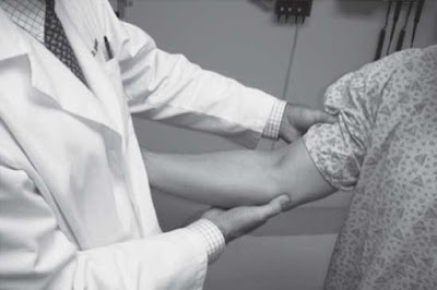
Physical exam should begin with inspection of the elbow to detect the presence of an effusion. If a UCL injury is suspected, the ligament should be palpated along its anatomic course from the medial epicondyle (origin) to the sublime tubercle (insertion) located on the ulna. Any tenderness within this area can represent significant injury to the ligament or the flexor-pronator musculature. Both passive and active range of motion should be compared with the opposite extremity, as it is common to observe slight elbow flexion contractures in the dominant extremity. Several physical examination tests can be used to specifically test the integrity of the UCL. The valgus stress test is performed with the arm stabilized against the examiner and the elbow flexed to 30 degrees ( Figure 24–1 ). Valgus stress is then gently applied at the elbow to detect any abnormal widening at the ulnohumeral joint and/or reproduction of painful symptoms. The “milking maneuver” is performed with the elbow flexed beyond 90 degrees, as the contralateral arm is used to grasp the thumb and generate a valgus force at the elbow ( Figure 24–2 ). If the patient reports pain with this test, a UCL injury should be suspected. The moving valgus stress test is performed with the examiner applying a valgus torque at the elbow in full flexion and quickly extending the elbow to produce a shear force ( Figures 24–3A and 24–3B ). This test is highly sensitive (100%) and specific (75%) for UCL injury.
Figure 24–1. The valgus stress test is performed with the arm stabilized against the examiner and
the elbow flexed to 30 degrees. Valgus stress is then gently applied at the elbow to detect any
abnormal widening at the ulnohumeral joint and/or reproduction of painful symptoms. (Courtesy of
Christopher Dodson, MD)
Figure 24–2. The “milking maneuver” is performed with the elbow flexed beyond 90 degrees, as the
contralateral arm is used to grasp the thumb and generate a valgus force at the elbow. If the patient
reports pain with this test, a UCL injury should be suspected. (Courtesy of Christopher Dodson, MD)
Figure 24–3. The moving valgus stress test is performed with the examiner (A) applying a valgus
torque at the elbow in full flexion and (B) quickly extending the elbow to produce a shear force.
This test is highly sensitive (100%) and specific (75%) for UCL injury. (Courtesy of Christopher
Dodson, MD)
Several basic and advanced imaging studies can be used to facilitate accurate diagnosis and guide proper treatment in any athlete who presents with a suspected UCL tear. Routine diagnostic imaging should begin with standard AP and lateral radiographs of the elbow. A “throwers series,” which includes two oblique radiographic views in internal and external rotation and an oblique axial view with the elbow in 110 degrees of flexion, can also be obtained to look for subtle associated pathology (ie, osteophyte formation, radiocapitellar osteochondritis dissecans, intraarticular loose bodies). Lastly, stress AP radiographs (x-rays obtained with valgus stress applied to the elbow) of both the injured and uninjured elbows can be used to detect any subtle differences in ulnohumeral widening secondary to UCL disruption. Advanced imaging, such as magnetic resonance imaging (MRI), can help identify ligament thickening from chronic overuse injury or more obvious full-thickness tears. Magnetic resonance arthrography obtained with intraarticular injection of gadolinium can aid in the diagnosis of partial undersurface tears of the ligament.
Some UCL tears can successfully be managed with a structured rehabilitation protocol. Early phases of rehabilitation include strict rest from all throwing activities and reduction of pain and inflammation with ice and nonsteroidal antiinflammatory drugs (NSAID). Gradual flexor-pronator strengthening exercises are incorporated, along with a progressive throwing program once all painful symptoms have resolved.
Surgical reconstruction of the UCL is indicated when patients demonstrate continued symptoms after a trial of structured rehabilitation. UCL reconstruction should only be performed in high-demand, throwing athletes who are willing to participate in an extensive postoperative rehabilitation program. When indicated, reconstruction involves securing a tendon graft to the humerus and ulna. Several techniques have been described, all of which provide good to excellent results in approximately 85% of cases. Common techniques used include the modified Jobe technique and the docking technique. It is clear that a muscle-splitting approach and minimal surgical manipulation of the ulnar nerve can limit postoperative morbidity and complications.
The main complications to beware of include damage to the ulnar or medial antebrachial cutaneous nerve during surgical dissection, ulnar or epicondylar fracture during tunnel drilling, and postoperative elbow stiffness from inadequate rehabilitation. In general, athletes can expect to return to competitive throwing 12 months after surgery if the shoulder, elbow, and forearm are pain-free and full range of motion has been restored.
COMPREHENSION QUESTIONS
24.1 A 19-year-old college javelin thrower presents with medial elbow pain during overhead throwing. He is diagnosed with a UCL tear after physical examination and MRI. What anatomic structure is the primary stabilizer to valgus stress at the elbow?
A. Anterior bundle of the UCLB. Transverse ligamentC. Radial headD. Flexor pronator mass
24.2 A 17-year-old baseball pitcher presents with medial elbow pain during overhead throwing. He is diagnosed with a UCL tear after physical examination and MRI. During what phase of throwing is he most likely to complain of medial elbow pain?
A. Follow-throughB. DecelerationC. AccelerationD. Wind-up
24.3 A 21-year-old javelin thrower is diagnosed with a full-thickness UCL tear. On presentation, he complains of numbness and tingling in the ring and small fingers. Which muscle group would he be most likely to have motor weakness in as well?
A. BrachioradialisB. Extensor carpi ulnarisC. Abductor digiti minimiD. Palmaris longus
ANSWERS
24.1 A. The anterior bundle of the UCL is the primary stabilizer to valgus stress at the elbow.
24.2 C. Throwing athletes typically complain of medial elbow pain during the acceleration phase of throwing. Valgus force generated at the elbow during the throwing motion is highest during the acceleration phase.
24.3 C. The patient is experiencing ulnar nerve paresthesias. If the patient had associated motor symptoms, they would manifest as weakness within the abductor digiti minimi. The other answer choices are not muscles innervated by the ulnar nerve.
CLINICAL PEARLS
|
► The anterior bundle of the UCL is the primary stabilizer to valgus stress at the elbow. ► Patients with UCL injuries typically report medial elbow pain during the acceleration phase of throwing. ► The most sensitive and specific clinical test for UCL injury is the moving valgus stress test. ► A muscle-splitting surgical approach and minimal manipulation of the ulnar nerve can limit postoperative morbidity to the flexor pronator mass and the ulnar nerve. ► Ulnar nerve transposition can be performed in conjunction with UCL reconstruction if patients demonstrate significant pathology related to the nerve (ulnar nerve subluxation or motor weakness). |
REFERENCES
Fleisig GS, Andrews JR, Dillma CJ, Escamilla RF. Kinetics of baseball pitching with implications about injury mechanisms. Am J Sports Med. 1995;23:233-239.
Hariri S, Safran MR. Ulnar collateral ligament injury in the overhead athlete. Clin Sports Med. 2010;29:619-644.
Jobe FW, Stark H, Lombardo SJ. Reconstruction of the ulnar collateral ligament in athletes. J Bone Joint Surg [Am]. 1986;68A:1158-1163.
Williams RJ, Urquhart ER, Altchek DW. Medial collateral ligament tears in the throwing athlete. Instr Course Lect. 2004;53:579-586.


0 comments:
Post a Comment
Note: Only a member of this blog may post a comment.