Ovarian Cancer (Epithelial) Case File
Eugene C. Toy, MD, Patti Jayne Ross, MD, Benton Baker III, MD, John C. Jennings, MD
CASE 59
A 62-year-old parous woman complains of a 3-month history of weight loss, abdominal bloating, unable to eat much without “indigestion.” She has no prior medical problems. On examination, her blood pressure is 110/60 mm Hg, heart rate is 80 beats per minute, and she is afebrile. She is alert and in no distress. Her heart and lung exams are normal. Her abdomen is moderately protuberant, and nontender and with normal bowel sounds. There is a fluid wave present. Based on the clinical findings, the physician suspects a gynecologic malignancy.
» What is the most likely diagnosis?
» What is likely to be best management of this patient?
ANSWER TO CASE 59:
Ovarian Cancer (Epithelial)
Summary: A 62-year-old parous woman with no medical history has a 3-month history of weight loss, abdominal bloating, and early satiety. She has a protuberant abdomen and a fluid wave. A gynecologic malignancy is suspected.
- Most likely diagnosis: Epithelial ovarian cancer.
- Management of this patient: Surgical staging and ovarian cancer debulking, followed by combination chemotherapy.
- Know that epithelial ovarian cancer presents abdominal bloating and indigestion, and is associated with ascites.
- Know that benign cystic teratomas (dermoid cysts) are the most common ovarian tumors in women younger than 30 years.
- Know that surgical therapy and cancer debulking followed by combination chemotherapy is the treatment of choice for epithelial ovarian cancer.
- Understand how to evaluate and manage adnexal masses in the various age groups.
Considerations
This 62-year-old woman has a 3-month history of weight loss, abdominal bloating, and indigestion. She is noted to have a protuberant abdomen which shows a fluid wave, which is most likely ascites. Based on the clinical presentation, the physician suspects a gynecologic malignancy. The presence of ascites would be consistent with epithelial ovarian cancer. The treatment of choice for epithelial ovarian cancer is surgical staging and debulking. Staging includes a total hysterectomy, bilateral salpingo-oophorectomy, omentectomy, lymph-node sampling, pelvic washings, and peritoneal biopsies. After maximal surgical debulking, the patient is usually treated with combination chemotherapy, such as with a paclitaxel and platinum combination. Endometrial cancer is the most common gynecologic malignancy, and usually presents with postmenopausal vaginal bleeding. Cervical cancer is the second most common gynecologic malignancy and classically presents with abnormal vaginal bleeding and/ or a cervical mass. Ovarian cancer is the third most common malignancy but the leading cause of gynecologic cancer death, principally due to discovery at a late stage. Vulvar cancer presents with itching, and/ or a labial ulcer or mass.
APPROACH TO:
Adnexal Masses and Ovarian Cancer
DEFINITIONS
CYSTIC TERATOMA (DERMOID CYST): A germ cell tumor that may contain all three germ cell layers. These are almost always benign (mature), although rarely they can be malignant (immature).
STRUMA OVARII: Benign cystic teratoma containing thyroid tissue, which can cause symptoms of hyperthyroidism.
OVARIAN NEOPLASM: An abnormal growth (either benign or malignant) of the ovary; most will not regress.
EPITHELIAL OVARIAN TUMOR: Neoplasm arising from the outer layer of the ovary, which can imitate the other epithelium of the gynecologic or urologic system. This is the most common type of ovarian malignancy, usually occurring in older women.
GERM CELL OVARIAN TUMOR: Neoplasm of the ovary derived from germ cells. This is the second most common type of ovarian neoplasm, occurring in young women.
SEX CORD-STROMAL TUMORS: Neoplasm of the ovary derived from the sex cords and supporting stroma of the ovary.
FUNCTIONAL OVARIAN CYST: Physiologic cysts of the ovary, which occur in reproductive-aged women, of follicular, corpus luteal, or theca lutein in origin.
CLINICAL APPROACH
Germ Cell Tumors
Germ cell tumors (Table 59– 1) represent about one-quarter of all ovarian tumors, and are the second most frequent type of ovarian neoplasms. They are found mainly in young women, usually in the second and third decades of life. The most common germ cell tumor is the benign cystic teratoma (dermoid). A germ cell malignancy usually presents as a pelvic mass and causes pain due to its rapidly enlarging size. Because of these symptoms, 60% to 70% of patients present as stage I, limited to one or both ovaries.

Teratomas
Mature (benign) cystic teratomas constitute over 95% of all ovarian teratomas. They make up 15% to 25% of all ovarian tumors, especially in the second and third decades of life. Teratomas contain tissues of all three embryonic layers, including endoderm, mesoderm, and ectoderm. The most common elements are ectodermal derivatives such as skin, hair follicles, and sebaceous or sweat glands. Although most are unilateral, they can appear bilaterally 10% to 15% of the time. Ultrasound features of dermoid cysts include a hypoechoic area or echoic band-like strand in a hypoechoic medium or the appearance of a cystic structure with a fat fluid level. Ultrasound is generally very accurate in the diagnosis of dermoid cysts. Torsion is the most frequent complication, with severe acute abdominal pain as the typical presenting symptom. This is more commonly seen during pregnancy, the puerperium, and in children or younger patients. Rupture is an uncommon complication and may present as shock or hemorrhage. A chemical peritonitis can be caused by the spill of the contents of the tumor into the peritoneal cavity. The treatment is usually a cystectomy or unilateral oophorectomy with inspection of the contralateral ovary.
Immature (malignant) teratomas contain all three germ layers, as well as immature or embryonal structures. They are uncommon and comprise less than 1% of ovarian cancers. They occur primarily in the first and second decades of life and are rare after menopause. Malignant teratomas contain immature neural elements and that quantity alone determines the grade. They are almost always unilateral. The prognosis is directly related to the stage and the grade or degrees of cellular immaturity. The treatment is a unilateral salpingo-oophorectomy with excision or extensive sampling of peritoneal implants. If the primary tumor is grade 1, no further treatment is warranted. However, if the primary tumor is grade 2 or 3 and if there are implants or recurrences, combination chemotherapy is usually indicated.
Struma Ovarii
Struma ovarii is a teratoma in which thyroid tissue is a major or exclusive element. They are usually unilateral, occurring more frequently in the right adnexa, and generally measure less than 10 cm in diameter. Preoperative clinical or radiologic diagnosis is very difficult. On magnetic resonance imaging, these tumors appear as complex multilobulated masses with thick septa, thought to represent multiple large thyroid follicles. Most of these tumors are benign, but about 10% can have malignant changes. They will rarely produce sufficient thyroid hormone to induce hyperthyroidism, with less than 5% leading to thyrotoxicosis. The treatment is usually cystectomy or salpingo-oophorectomy.
Epithelial Tumors
Epithelial tumors (Table 59– 2) represent about two-thirds of all ovarian tumors, and are the most frequent type of ovarian neoplasms, typically occurring in women over 30 years.
The serous subtype is most common and more often bilateral. Mucinous tumors are characterized by their large size, and if ruptured, may lead to pseudomyxomaperitonei, a condition in which the mucinous material spills out into the intra-abdominal cavity. This can lead to repeat bouts of bowel obstruction. Endometrioid tumors of the ovary may coexist with a primary endometrial carcinoma of the uterus.

Epithelial Ovarian Cancer
Epithelial ovarian cancer is the most common cause of gynecologic cancer death and fifth most frequent cause of cancer death in women. Approximately 5% to 10% of cases are familial with first-degree relatives having ovarian cancer or ovarian– breast cancer; some of these involve the BRCA 1 or 2 mutation. Most patients have widespread metastasis at the time of diagnosis because of the early spread of the malignant cells to peritoneal and bowel surfaces. Gastrointestinal symptoms such as bloating, early satiety, increased abdominal girth, or abdominal pain are frequently ignored, but noted in retrospect.
The physical examination may show an abdominal or pelvic mass, ascites, or omental nodules. The tumor marker cancer antigen (CA)-125 is elevated in most epithelial ovarian tumors. CA-125 is more specific in postmenopausal women, since a variety of diseases during the reproductive years can elevate the CA-125 level. Imaging is important to characterize the pelvic mass. Malignant ascites is common with cancer, as it is spread to the small bowel, omentum, and lymphatics.
The treatment of epithelial tumors is surgical, and if malignancy is confirmed, cancer staging is indicated. Treatment of epithelial ovarian cancer involves a combination of surgical staging with maximum removal of the tumor (debulking) and combination chemotherapy especially with a platinum agent (cisplatinum or carboplatinum) and a taxane.
Sex Cord-Stromal Tumors
Sex cord-stromal (Table 59– 3) represents approximately 6% of ovarian neoplasms and the majority of hormonally active neoplasms. Granulosa cell tumors contain functional granulosa cells which produce estrogen and supporting theca cells. Approximately 5% of these tumors occur before puberty and can lead to precocious puberty. Sertoli–Leydig tumors secrete androgens which can lead to masculinization and hirsutism. Sex cord-stromal tumors appear solid on ultrasound, with fibromas being the most common benign solid neoplasm of the ovary.
Adnexal Masses
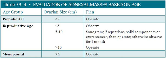
The evaluation of adnexal masses is guided by the suspicion of neoplasm (benign or malignant). At the extremes of ages, there are few functional ovarian cysts and the management is straightforward (Table 59– 4).
During the reproductive years, functional ovarian cysts, such as follicular and corpus luteal cysts, sometimes make the evaluation difficult. In general, any adnexal mass greater than 10 cm in size is likely to be a tumor and should be excised. Any adnexal mass less than 5 cm in size suggests a functional cyst. Between 5 and 10 cm, the sonographic features may help to distinguish functional versus neoplasm. Septations, solid components, or excrescences (growth on surface or inner lining) are consistent with a neoplastic process, whereas a simple cyst is more suggestive of a functional cyst. Sometimes, a practitioner will choose to observe and repeat imaging in an adnexal mass that is between 5 and 10 cm in size and operate if it is persistent.
Emerging Concepts
Over the past several years, the proposed etiology of several types of epithelial ovarian cancer (serous, endometrioid, clear cell) is speculated to arise from the fallopian tube, particularly the fimbriated end. These seem to be associated with high grade and undifferentiated malignancies. Many experts recommend salpingectomy at the time of hysterectomy as a means of reducing ovarian cancer risk. Regarding therapy, several randomized trials have shown increased survival with the combination of intraperitoneal and intravenous (IV) chemotherapy versus only IV chemotherapy for epithelial ovarian cancer. There seems to be more adverse complications with intraperitoneal chemotherapy.

|
CASE CORRELATION - See also Case 36 (Pelvic Inflammatory Disease), Case 40 (Uterine Fibroids), Case 53 (Sertoli–Leydig Cell Tumor of the Ovary), and Case 56 (Endometriosis) for a differential diagnosis of pelvic masses:
- Uterine fibroids—typically midline and irregular.
- Tubo-ovarian abscess—typically with some adnexal tenderness.
- Sertoli–Leydig cell tumor—usually androgen effects.
- Endometrioma—usually less than 8 cm, associated with dysmenorrhea and dyspareunia.
|
COMPREHENSION QUESTIONS
59.1 A 5-year-old girl is noted to have breast enlargement, vaginal bleeding, and an 8-cm pelvic mass. Which of the following is the most likely etiology?
A. Benign cystic teratoma (dermoid)
B. Endodermal sinus tumor
E. Granulosa-theca cell tumor
59.2 A 25-year-old woman is noted to have a 4-cm simple cyst of the right ovary. She denies any abdominal pain, nausea, or vomiting. Which of the following is the next best step?
C. Exploratory laparotomy
59.3 Which of the following is the best treatment for a suspected dermoid cyst found in an 18-year-old nulliparous woman?
A. Total abdominal hysterectomy
B. Unilateral salpingo-oophorectomy
Match the following sonographic findings (A-D) with the ovarian tumor type (59.4-59.6)?
D. Ascites is commonly seen
59.4 Granulosa cell tumor
59.5 Benign cystic teratoma (dermoid cyst)
59.6 Follicular cyst
59.7 A 44-year-old woman is noted to have a 30-cm tumor of the ovary. Which of the following is the most likely cell type?
ANSWERS
59.1 E. This is a young child with precocious puberty, which suggests an estrogensecreting tumor. This is most likely a granulosa-theca cell tumor, best treated by surgery. These are sex cord-stromal tumors.
59.2 A. When the ovarian cyst in the reproductive-aged female is less than 5 cm in diameter, the most likely cause is a physiologic cyst such as a follicular cyst or corpus luteum. Expectant management and reassessment in 1 to 3 months is the best next step.
59.3 C. Ovarian cystectomy is the best treatment for benign cystic teratomas in a younger patient especially when future childbearing is desired. Because of the 15% to 20% risk of bilaterality, the contralateral ovary should be inspected for a possible dermoid.
59.4 A. Granulosa cell tumors and Sertoli–Leydig cell tumors are usually solid on ultrasound, and may secrete sex hormones. Typically, granulosa-theca cell tumors produce estrogens, whereas Sertoli–Leydig cell tumors make androgens.
59.5 C. Benign cystic teratomas (dermoid cysts) are complex cysts since they usually have both solid and cystic components. The best treatment of a dermoid in a young woman is ovarian cystectomy. Ovarian torsion is the most frequent complication. Immature teratomas contain all three germ layers, as well as immature or embryonal structures. Malignant teratomas contain immature neural elements, and the grade of the tumor is determined by the amount of neural tissue involved.
59.6 B. Follicular cysts are generally simple cysts without septations or solid parts. They are among the physiologic cysts of the ovary, which occur in reproductive- aged women. Other physiologic, or functional, cysts include corpus luteal or theca lutein cysts.
59.7 D. Mucinous tumors of the ovary can grow to be very large. If they rupture intra-abdominally, they may cause pseudomyxomaperitonei, which leads to repeated bouts of bowel obstruction. They are of epithelial origin. The most common type of epithelial ovarian tumor is the serous type, which unlike the mucinous tumors, usually occurs bilaterally. The tumor marker CA-125 is elevated in most epithelial ovarian tumors and is more specific in postmenopausal women because a variety of diseases that occur during the reproductive years can show an elevated CA-125 level.
CLINICAL PEARLS
» The most common ovarian tumor in a woman younger than 30 years is a benign cystic teratoma (dermoid cyst). The best treatment of a dermoid in a young woman is ovarian cystectomy.
» The most common ovarian tumor in a woman older than 30 years is epithelial in origin, most commonly serous cystadenoma.
» An ovarian mass larger than 5 cm in a postmenopausal woman most likely represents an ovarian tumor and should generally be removed. An ovarian mass that is larger than 2 cm in a prepubertal girl likewise should be investigated and usually requires removal.
» During the reproductive years, functional ovarian cysts are common and are usually smaller than 5 cm in diameter. Any ovarian cyst larger than 10 cm in a reproductive-aged woman is probably a neoplasm and should be excised.
» The tumor marker CA-125 is elevated in most epithelial ovarian cancers. It is more specific in postmenopausal women.
» Mucinous tumors of the ovary can grow to be very large. If they rupture intra-abdominally, they may cause pseudomyxomaperitonei, which can lead to repeated bouts of bowel obstruction.
» Ascites is a common sign of ovarian malignancy.
» Ovarian cancer staging consists of total hysterectomy, bilateral salpingooophorectomy, omentectomy, peritoneal biopsies, peritoneal washings or sampling of ascitic fluid, and lymphadenectomy.
» After maximum debulking of the epithelial cancer, combination chemotherapy with a platinum agent and a taxane is used.
|
REFERENCES
American College of Obstetricians and Gynecologists. Management of adnexal masses. ACOG Practice
Bulletin 83. Washington, DC; July 2007 (Reaffirmed 2015).
American College of Obstetricians and Gynecologists. Salpingectomy for Ovarian Cancer Prevention.
ACOG Committee Opinion 620; Jan 2015.
Coleman RL, Gershenson DM. Neoplastic diseases of the ovary. In: Katz VL, Lentz GM, Lobo RA,
Gersenson DM, eds. Comprehensive Gynecology. 6th ed. St. Louis, MO: Mosby-Year Book; 2012:
839-882.
Katz VL. Benign gynecologic lesions. In:
Katz VL, Lentz GM, Lobo RA, Gersenson DM, eds. Comprehensive
Gynecology. 6th ed. St. Louis, MO: Mosby-Year Book; 2012:419-471.
National Cancer Institute. Ovarian epithelial cancer treatment. http:/ / www.cancer.gov/ cancertopics/
pdq/ treatment/ ovarianepithelial/ HealthProfessional/ page1; Accessed 1.10.2015.
Nelson AL, Gambone JC. Congenital anomalies and benign conditions of the ovaries and fallopian
tubes. In: H acker NF, Gambone JC, Hobel CJ, eds. Essentials of Obstetrics and Gynecology. 6th ed.
Philadelphia, PA: Saunders; 2015:248-255.
Stany MP, Hamilton CA. Benign disorders of the ovary. Obstet Gynecol Clin North Am. 2008;35(2):
271-284.


0 comments:
Post a Comment
Note: Only a member of this blog may post a comment.