Pleural Effusion, Parapneumonic Case File
Eugene C. Toy, MD, Gabriel M. Aisenberg, MD
Case 17
A 32-year-old woman presents to the emergency center complaining of a productive cough, fever, and chest pain for 4 days. She was seen 2 days ago in her primary care provider’s clinic with the same complaints; she was diagnosed clinically with pneumonia and was sent home with oral azithromycin. Since then, her cough has diminished in quantity. However, the fever has not abated, and she still experiences left-sided chest pain, which is worse when she coughs or takes a deep breath. In addition, she has started to feel short of breath when she walks around the house. She has no other medical history. She does not smoke and has no history of occupational exposure. She has not traveled outside of the United States and has no sick contacts.
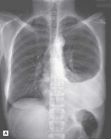
On physical examination, her temperature is 103.4 °F, heart rate is 116 beats per minute (bpm), blood pressure is 128/69 mm Hg, and respiratory rate is 24 breaths/min and shallow. Her pulse oximetry is 94% saturation on room air. Physical examination is significant for decreased breath sounds in the lower half of the left lung fields posteriorly, with dullness to percussion between the fifth and eighth intercostal spaces at the midclavicular line. There are a few inspiratory crackles in the midlung fields, and her right lung is clear to auscultation. She has sinus tachycardia with no murmurs. She has no cyanosis. Figure 17–1 shows her chest x-ray films.
▶ What is the most likely diagnosis?
▶ What is your next step in management?
▶ What are the most common causes of this condition?
Figure 17–1. (A) Posteroanterior film of the chest. (B) Lateral chest film of the same patient. (Courtesy of Dr. Jorge Albin.)
ANSWERS TO CASE 17:
Pleural Effusion, Parapneumonic
Summary: A 32-year-old previously healthy woman presents with
- A 4-day history of productive cough, fever, and chest pain
- Clinical diagnosis of community-acquired pneumonia, not improved with oral azithromycin
- Vital signs: febrile, tachycardic, tachypneic with hypoxemia on room air
- Diminished breath sounds and left-sided dullness to percussion
- Chest radiography confirming a large left-sided pleural effusion
- Effusion that is likely caused by infection in the adjacent lung parenchyma and that may be the cause of her failure to improve on antibiotics
Most likely diagnosis: Parapneumonic effusion as a complication of pneumonia.
Next step: Diagnostic thoracentesis to help diagnose the cause of the pleural effusion and to determine the necessity for fluid drainage.
Most common causes: Underlying pneumonia, malignancy, pulmonary embolism, tuberculosis.
- Understand the use of Light criteria to distinguish transudative effusions from exudative effusions as a guide to the etiology of the effusion. (EPA 1, 3)
- Describe the pleural fluid characteristics that suggest a complicated parapneumonic effusion or empyema and the need for drainage. (EPA 3, 4)
- State the treatment of a complicated parapneumonic effusion that does not improve after thoracentesis. (EPA 4, 10)
Considerations
If the pleural effusion is large and free flowing, which can be evaluated with a lateral decubitus film, then diagnostic thoracentesis can easily be accomplished. It is important to determine if the effusion is, in fact, caused by the pneumonia, and, if so, whether it is likely to resolve with antibiotics alone or will require drainage with tube thoracostomy.
APPROACH TO:
Pleural Effusion
DEFINITIONS
EMPYEMA: Collection of pus in the pleural cavity.
EXUDATE: Effusion caused by inflammatory or malignant causes, with high protein or high lactate dehydrogenase (LDH) levels.
PLEURAL EFFUSION: Accumulation of fluid in the pleural space.
TRANSUDATE: Effusion caused by alteration of hydrostatic and oncotic forces, with low protein and low LDH levels.
CLINICAL APPROACH
Pathophysiology
Pleural effusions occur in 40% of patients with an underlying bacterial pneumonia. Most of these effusions should resolve with appropriate antibiotic treatment, but if the fluid characteristics predict a “complicated” parapneumonic effusion, urgent tube drainage is indicated to prevent formation of fibrous peels, which may need surgical decortication.
Diagnostic thoracentesis should be considered for every patient who presents with a pleural effusion with unknown cause. An exception to this rule is if the patient is known to have heart failure (HF) with equal bilateral effusions or if the effusion is too small—that is, less than 10 mm—on lateral decubitus film. However, if the pleural effusion of HF does not significantly improve after a trial of diuresis, a diagnostic thoracentesis should be performed. Another exception would be in patients with fluid overload states, such as in end-stage renal disease or nephrotic syndrome, where dialysis will help with the clearing of effusions. Table 17–1 gives

the correlations of pleural fluid appearance. As little as 5 to 10 mL can be visualized on a lateral decubitus film (it is more reliable in detecting smaller effusions), and fluid volume more than 500 mL usually obscures the whole hemidiaphragm. Ultrasound is an additional tool that can be easily used to identify pleural effusions.
Transudate Versus Exudate. Transudative and exudative fluids can be differentiated by the amount of measured protein and LDH, which correlate with the pathophysiology of the fluid formation. Approximately 12 mL of pleural fluid is formed every day by the parietal pleural capillaries and absorbed also by the parietal pleura (lymphatics). Processes that disturb this equilibrium lead to fluid accumulation. Clinical settings in which the hydrostatic pressure is increased (HF and constrictive pericarditis), the oncotic pressure is decreased (nephrotic syndrome and cirrhosis), or the intrapleural pressure is reduced (atelectasis), lead to the formation of a “transudate.” In contrast, “exudates” are more a result of local inflammation and increased capillary permeability—for example, infection, malignancy, and connective tissue diseases, which cause proteins to leak into the pleural space. Less commonly, impaired lymphatic drainage, as occurs in chylothorax or lymphangitic spread of a malignancy, may cause an exudative fluid. Pulmonary emboli can cause both exudative and transudative effusions (exudative is much more common). Tables 17–2 and 17–3 list the etiologies of transudative and exudative pleural effusions, respectively.
Light Criteria. The most widely used criteria to distinguish between a transudative and exudative fluid are the Light criteria first described in 1997. For a fluid to be labeled an exudate, it must meet at least one of the following criteria (transudates meet none of these criteria):
- Pleural fluid protein/serum protein ratio > 0.5
- Pleural fluid LDH/serum LDH ratio > 0.6
- Pleural fluid LDH > 2/3 the upper limit of normal for serum LDH
Pleural LDH correlates with the degree of pleural inflammation and, along with fluid protein, should always be sent in the initial evaluation.
Treatment
In contrast to the simple “diagnostic” thoracentesis that can be performed to evaluate the contents of the fluid, a “therapeutic” thoracentesis may also be performed if the patient is dyspneic as a result of a significant amount of fluids. The purpose is to remove large volumes of fluid (up to 1.5 L) for patient comfort and clinical improvement. Of note, removal of large amounts of fluid puts the patient at risk for developing reexpansion pulmonary edema.
If tube thoracostomy drainage is required, a chest tube is placed until the drainage rate has decreased to less than 50 mL/d. Postdrainage imaging must be obtained to confirm complete drainage of fluid and to assess the need for placement of a second tube if the fluid has not been adequately drained (as is often seen if the effusion is loculated). The following fluid characteristics suggest the need for chest tube drainage:
- Most sensitive: pH < 7.20 (normal pH 7.6)
- Positive Gram stain or culture of fluid
- Presence of loculations
- Empyema (frank pus in the pleural space)
- Glucose less than 60 mg/dL
- LDH more than 1000 U/L
If the patient does not meet the criteria for immediate drainage, a 1-week trial of antibiotics is indicated, with close reevaluation of those patients who do not respond or who clinically deteriorate.
In empyema, 4 to 6 weeks of antibiotic therapy are necessary for complete sterilization. Poorly draining or multiloculated empyemas are treated further by administering a combination of fibrinolytic agents such as tissue plasminogen activator (tPA) and deoxyribonuclease (DNase) through the chest tube. Video-assisted thoracoscopic surgery with debridement and drainage is the next option if the combination of tPA and DNase fail in clearing the loculations. Thus, surgical referral is encouraged within a week of failure of medical therapy (tube thoracostomy with or without tPA and DNase).
CASE CORRELATION
- See also Case 14 (Pulmonary Embolism), Case 15 (Chronic Obstructive Pulmonary Disease), and Case 16 (Chronic Cough/Asthma).
COMPREHENSION QUESTIONS
17.1 A 55-year-old man with HF presents to the emergency department with a 1-week history of dyspnea on exertion and swelling in both ankles. He has had no fever or cough. Chest radiography shows bilateral pleural effusions. Which of the following is the most likely pleural fluid characteristic if thoracentesis is performed?
A. Pleural fluid LDH 39, LDH ratio 0.2, protein ratio 0.7
B. Pleural fluid LDH 39, LDH ratio 0.2, protein ratio 0.1
C. Pleural fluid LDH 599, LDH ratio 0.9, protein ratio 0.1
D. Pleural fluid LDH 599, LDH ratio 0.9, protein ratio 0.7
17.2 A 39-year-old man develops a moderate free-flowing pleural effusion following a left lower lobe pneumonia. Thoracentesis reveals straw-colored fluid with gram-positive diplococci on Gram stain, pH 6.9, glucose 32 mg/dL, and LDH 1890. Which of the following is the best next step?
A. Send the fluid for culture.
B. Continue treatment with antibiotics for pneumococcal infection.
C. Drain the effusion via tube thoracostomy.
D. Schedule a follow-up chest x-ray in 2 weeks to document resolution of the effusion.
17.3 A 69-year-old man is being seen in the emergency center for gradually worsening dyspnea and a nagging cough over the past 3 months. He denied feeling warm or having fever. The chest radiograph shows a right-sided pleural effusion. A diagnostic thoracentesis reveals gross blood in the fluid. Which of the following is the most likely diagnosis?
A. Parapneumonic effusion
B. Malignancy in the pleural space
C. Rupture of aortic dissection into the pleural space
D. Pulmonary embolism with pulmonary infarction
ANSWERS
17.1 B. Heart failure is commonly associated with bilateral pleural effusions, which are transudative, as a consequence of alteration of Starling forces. The effusions of heart failure are best managed by treating the heart failure, for example, with diuretics, and typically do not require thoracentesis. Per Light criteria, the other answer choices would be classified as an exudative pleural effusion. Exudative effusion criteria include pleural fluid: serum protein > 0.5; pleural fluid: serum LDH > 0.6; and pleural fluid LDH > 2/3 upper limits for serum.
17.2 C. Drainage with chest thoracostomy is the best treatment for this patient. The positive Gram stain, low pH, low glucose, and markedly elevated LDH all suggest that this parapneumonic effusion is “complicated,” that is, it is unlikely to resolve with antibiotic therapy; this effusion is likely to produce loculated pockets of pus, which will require drainage with tube thoracostomy. While continuing the therapy with antibiotics (answer B) is appropriate, it is also insufficient in the case of an empyema, which is the situation at present with this patient. Answer A (Send the fluid for culture) is usually performed, but it often is negative for growth; although antibiotics and culture are important adjuvant measures, the most important intervention is fluid drainage. Answer D (Schedule a follow-up chest x-ray in 2 weeks) is inappropriate and will lead to patient decompensation.
17.3 B. The most common causes of hemorrhagic pleural effusion are trauma, malignancy, and pulmonary embolism. Pulmonary embolism (answer D) would be suggested by acute onset of dyspnea and pleuritic chest pain rather than this subacute presentation. Aortic rupture (answer C) can produce a hemothorax but would have an acute presentation with chest and back pain and hemodynamic compromise. Parapneumonic effusion (answer A) is a collection of purulent fluid that arises from pneumonia, lung abscess, or bronchiectasis; the aspirated fluid would be purulent and not bloody.
CLINICAL PEARLS
▶ Transudative effusions meet none of the following criteria (exudative effusions meet at least one): (a) pleural fluid protein/serum protein ratio more than 0.5; (b) pleural fluid LDH/serum LDH ratio more than 0.6; (c) pleural fluid LDH greater than two-thirds normal serum LDH.
▶ Tube thoracostomy or more aggressive drainage of parapneumonic effusion usually is required with gross pus (empyema), positive Gram stain or culture, glucose less than 60 mg/dL, pH less than 7.20, and loculations.
▶ The most common cause of pleural effusion is HF, which typically results in bilateral symmetric transudative effusions and is best treated with diuresis.
▶ The most common causes of bloody pleural effusion (in the absence of trauma) are malignancy and pulmonary embolism with infarction.
REFERENCES
Colice GL, Curtis A, Deslauriers J, et al. Medical and surgical treatment of parapneumonic effusions: an evidence-based guideline. Chest. 2000;118:1158-1171.
Keane MP, Lynch JP. Pleuropulmonary manifestations of systemic lupus erythematosus. Thorax. 2000;55:159-166.
Light RW. Pleural effusion. N Engl J Med. 2002;346:1971-1977.
Light RW. Parapneumonic effusions and empyema. Proc Am Thorac Soc. 2006;3(1):75-80.
Light RW. Disorders of the pleura and mediastinum. In: Longo DL, Fauci AS, Kasper DL, et al, eds. Harrison’s Principles of Internal Medicine. 20th ed. New York, NY: McGraw Hill; 2018:2178-2182.
Shaw M, Collins BF, Ho LA, et al. Rheumatoid arthritis-associated lung disease. Eur Respir Rev. 2014;24(135):1-16.
Strange C. Management and prognosis of parapneumonic pleural effusion and empyema in adults. Broaddus VC, Ramirez JA, ed. UpToDate. Waltham, MA: UpToDate; 2019 https://www.uptodate
.com. https://www.uptodate.com/contents/management-and-prognosis-of-parapneumonic-pleural-effusion-and-empyema-in-adults. Accessed June 12, 2019.


0 comments:
Post a Comment
Note: Only a member of this blog may post a comment.