Basal Ganglia Case File
EUGENE C.TOY, MD, RAHUL JANDIAL, MD, PhD, EVAN YALE SNYDER, MD, PhD, MARTIN T. PAUKERT, MD
CASE 25
A 62-year-old male presents to his general practitioner complaining of a steady tremor in his hands, which has slowly progressed for the past 6 weeks. The patient also states that walking has become increasingly difficult although he attributes this to old age. Upon physical examination, the patient has increased muscle tone with a notably hunched posture and a resting tremor. When asked to make purposeful movements, the patient is slow to initiate the movement; however, the tremor is alleviated while moving. All cranial nerves are intact and reflexive examinations normal. The patient shows no symptoms of dementia, Alzheimer, or any other cognitive disorders. An MRI is ordered which shows mild brain atrophy appropriate for his age, but otherwise unremarkable. You make the diagnosis of Parkinson disease.
- What microscopic structures are found in the neurons of patients with this disorder?
- What are the treatment options available?
ANSWERS TO CASE 25: BASAL GANGLIA
Summary: A 62-year-old male with bradykinesia and a steady resting tremor in both hands. His tremor is temporarily mitigated while making purposeful movements. The patient has increased muscle tone and a hunched posture. When asked to walk, the patient’s gait is disturbed. The remainder of the physical examination is significant for normal reflexive responses and intact cranial nerves. A magnetic resonance image reveals mild global brain atrophy appropriate for the patient’s age, but is otherwise normal.
- Microscopic pathology: Lewy bodies are eosinophilic cytoplasmic inclusions with a halo of radial fibrils and are composed mainly of the protein alpha-synuclein. These pathologic structures are thought to accumulate over time and disrupt normal intracellular functions of nerve cells.
- Treatment options: While no current treatment options are curative, administration of levodopa (a precursor to Dopamine) and other Parkinsonism medications help alleviate the symptoms of Parkinson disease. Some patients may also be candidates for neurosurgical interventions such as thalamotomy, subthalamotomy, or pallidotomy procedures to alleviate movement symptoms once medical treatments have become ineffective. Also, current research shows that embryonic stem cell transplantation into the striatum may one day become an effective treatment option for patients with Parkinson disease.
CLINICAL CORRELATION
Parkinson disease is a common neurological disorder of the basal ganglia arising from neural cell degeneration within the pars compacta region of the substantia nigra. The loss of these pigmented dopaminergic neurons reduces the amount of dopamine synthesized by the substantia nigra. Without the appropriate amount of dopamine in the striatum, there is an antagonistic effect on the direct pathway and agonistic effect on the indirect pathway of the nigrostriatal projection. This often leads to hypokinesia. The direct pathway has the net effect of exciting thalamic neurons, which in turn make excitatory connections with cortical neurons. The indirect pathway has the net effect of inhibiting thalamic neurons, thereby inhibiting cortical neurons. The pathological cause of cell death in Parkinson disease is still unclear and the majority of cases are idiopathic. Other causes of Parkinson syndrome are related to drug-induced, toxic, genetic, and traumatic etiologies. Parkinsonism symptoms are also observed in individuals after longterm use of antipsychotic medications such as Haldol or after ingestion of the neurotoxin MPTP.
APPROACH TO BASAL GANGLIA
Objectives
- Know the anatomical structures and cellular projections of the basal ganglia.
- Be able to describe the different neurological functions of the basal ganglia.
- Know common disorders associated with the basal ganglia.
Definitions
Striatum: The caudate nucleus and the putamen. Together, the two nuclei act as the “receiving portion” of the basal ganglia.Pallidum: Comprised of the internal (GPi) and external (GPe) portion of the globus pallidus, the pallidum plays a modulatory role between the striatum and the thalamus. The efferent connections from the substantia nigra and the pallidum act as a mediator between the basal ganglia and the rest of the nervous system.Medium spiny neurons: The most common cell type found in the striatum. Contain the inhibitory neurotransmitter gamma-aminobutyric acid (GABA).Hemiballismus: Involuntary, violent movements of the contralateral side of the body resulting from lesions involving the subthalamic nucleus.Chorea: A movement disorder of the basal ganglia characterized by a rapid, irregular flow of motions as well as grimacing movements of the face.Athetosis: A movement disorder of the basal ganglia characterized by slow, writhing movements of a wormlike character involving the extremities, trunk, and neck.Disinhibition: Inhibition of an inhibiting projection pathway.
DISCUSSION
The basal ganglia are composed of the caudate nucleus, putamen, and globus pallidus. Because of the high degree of cellular connections with these three structures, the substantia nigra and subthalamic nucleus are also considered components of the basal ganglia. The basal ganglia have significant connections with both the thalamus and the cortex. It functions primarily in the modification and elaboration of movements initiated by the primary motor cortex. A lesion in one of the nuclei within the basal ganglia will therefore produce a disruption in movement and muscle tone but no pareses.
Other significant roles of the basal ganglia include cognitive functions such as verbal working memory, motor planning, repetitive movement learning, and the association of motivation and emotions to the execution of movements.
The caudate nucleus and the putamen together are termed the striatum. Together, these two nuclei act as the “receiving portion” of the basal ganglia.
The main afferent connections travel from the cerebral cortex, the intralaminar thalamic nuclei, and the dopamine-containing cell groups in the mesencephalon. The putamen largely receives information from the primary motor and somatosensory cortex, while the caudate nucleus receives the majority of its inputs from the association areas of the cortex. The intralaminar thalamic nuclei project cells to the striatum in large part from the centromedian nucleus. The dopaminergic afferent connections of the striatum arise from the substantia nigra and the ventral tegmental area.
The primary efferent pathways from the basal ganglia begin in the globus pallidus and substantia nigra and project to the thalamus, mesencephalic tegmentum, and superior colliculus. The efferent connections from the substantia nigra and the pallidum act as a mediator between the basal ganglia and the rest of the nervous system.
The globus pallidus is composed of internal and external segments, each receiving afferents input from the striatum and subthalamic nucleus. The actions of the striatal fibers on the internal segment of the globus pallidus (GPi) are inhibitory while the effects from the subthalamic nucleus are excitatory. The activity of the GPi is determined from the summation of inhibitory and excitatory input. The efferent output from the GPi acts on the substantia nigra and the thalamus. While the external portion of the globus pallidus (GPe) projects to the subthalamic nucleus, the substantia nigra has efferent connections with the thalamus and superior colliculus. The thalamus functions as a relay center for information projecting in both directions between the basal ganglia and the cerebral cortex. The basal ganglia additionally regulate muscle movements and tone through its efferent input to the reticulospinal tract and reticular formation. Six different cell types have been identified in the striatum, the most common of which are medium spiny neurons. These contain the inhibitory neurotransmitter GABA.
The substantia nigra has two components: the pars compacta and the pars reticula. The pars compacta is densely packed with neuromelanin-containing neurons (giving the nucleus its dark color). The afferent connections of the substantia nigra come from many different cell groups, the majority being from the striatum. The inhibitory afferents of the substantia nigra contain the neurotransmitter GABA. Excitatory afferents originate from the pedunculopontine nucleus and subthalamic nucleus transmitting glutamate, the raphe nucleus–transmitting serotonin, the locus coeruleus using norepinephrine, and the basal forebrain.
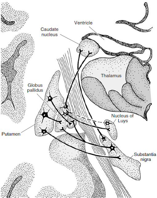
The efferent connections of the substantia nigra primarily project to the striatum and thalamus, with some association to the superior colliculus and reticular formation (see Fig. 25-1). The pars compacta contains the dopaminergic nigrostriatal neurons, making it a key location in Parkinson disease. The nigrostriatal pathway has the dual effect of exciting the direct pathway and inhibiting the indirect pathway—when this pathway is destroyed in Parkinson disease, then the indirect pathway cannot be inhibited. The pars reticula contains inhibitory GABAergic nigrothalamic neurons.
Figure 25-1. Diagram of the basal ganglia in the coronal plane, illustrating the main interconnections. (With permission from Adam and Victor’s Principles of Neurology. 7th ed. Figure 15-2, page 303.)
Dopamine is one of the key modulatory neurotransmitters in the striatum. Dopamine receptors can be divided into D1 and D2 types, each of which posses many of their own subtypes. The receptors influence many types of ion channels on both presynaptic and postsynaptic terminals. This allows the neurotransmitter to affect the stabilization of the membrane potential and contribute to keeping that potential in a range where the cell is apt to fire in bursts, that is, in a state suited for efficient signal transmission.
Excitatory afferent connections are received from the motor cortex by the subthalamic nucleus. The subthalamic nucleus sends the majority of its efferent connections to the pars reticula and the globus pallidus. The effects of the subthalamopallidal efferents are excitatory. The excitatory influences of the striatum determine the activity of the GABA-containing neurons within the GPi.
The subthalamic nucleus is believed to control or stop ongoing movements. Because of the inhibitory effects that the GPi has on the thalamocortical neurons, increased activity in the subthalamic nucleus would result in an inhibition of voluntary movement as a result of the inhibition of the motor cortex. The clinical presentation of hemiballismus, involuntary, violent movements of the contralateral side of the body resulting from lesions involving the subthalamic nucleus, may be because of increased activity of the thalamocortical neurons. Several diseases have been linked to the basal ganglia. Parkinson disease is a neurodegenerative disorder of the dopaminergic neurons of the substantia nigra. The disease is characterized by tremor, rigidity, akinesia, and postural instability. Current therapies for Parkinson disease are limited to the treatment of the movement symptoms.
Huntington disease is an inherited disease that results from progressive neurodegeneration of primarily GABAergic neurons within the striatum. It is characterized in its early stages by forgetfulness, depression, sudden falls, irritability, and choreiform movements which gradually progress until the patient is no longer ambulatory. In the late stages of the disease, continued cell deterioration eventually results in dementia. A locus for the disease has been identified on the short arm of chromosome 4 allowing carriers of the disease to be identified before symptoms present. The disease is caused by a selective loss of striatal neurons in the indirect pathway. Without the inhibition from the indirect pathway, the thalamic neurons can fire spontaneously, causing the motor cortex to execute motor programs with no control by the person. Tourette syndrome is a disorder involving a functional disturbance between the basal ganglia and the frontal cortex. The syndrome presents with multiple tics, often associated with uncontrollable vocal outbursts. Patients with Tourette syndrome are often treated with dopamine antagonists to relive the motor symptoms. Tardive Dyskinesia is an iatrogenic disorder that results from the long-term administration of neuroleptic medications such as haloperidol, chlorpromazine, and thioridazine. The disorder is characterized by uncontrollable movements of the head, face, lips, and tongue, and rigidity. The symptoms are caused by sensitization of dopamine receptors in the mesolimbic pathway. This sensitization results in an imbalance in communication between the nigrostriatal pathway and the basal nuclear motor loop.
COMPREHENSION QUESTIONS
Refer to the following case scenario to answer questions 25.1-25.3:
A 38-year-old man is brought into your office by his family because he has begun to behave somewhat strangely recently. He has been increasingly irritable, and withdrawn from activities he normally enjoys. Additionally, he has been having strange fidgeting movements of his hands and fingers nearly all the time that he did not have in the past. Based on this history and your physical examination findings, you perform an MRI, which is suggestive of Huntington disease, a disorder that affects the basal ganglia.
[25.1] With which of the following processes are the basal ganglia most associated?
A. Directly controlling input to the alpha motor neuronsB. Planning and execution of complex motor activitiesC. Modification of movements initiated by the motor cortexD. Integration and smoothing of multiple movements
[25.2] Which of the following structures serves as the primary input to the basal ganglia?
A. StriatumB. Globus pallidusC. Subthalamic nucleusD. Substantia nigra
[25.3] Which of the following structures serves as the primary output from the basal ganglia?
A. StriatumB. Internal segment of the globus pallidusC. External segment of the globus pallidusD. Subthalamic nucleus
Answers
[25.1] C. The primary function of the basal ganglia is thought to be modulation of cortical output, including modification of movements initiated by the motor cortex. The major outputs of the basal ganglia are inhibitory to the thalamus, and the variety of basal ganglia circuits alters these outputs to further inhibit or disinhibit the thalamus. Since the thalamic outputs to the cortex are for the most part excitatory, the basal ganglia are capable of inhibiting or disinhibiting the cortex. It is thought that these actions serve to somehow select the appropriate motor programs at the right time, while suppressing other programs that are not needed at the time. The primary motor cortex directly controls alpha motor neurons, the premotor and supplemental motor areas are involved in the planning of complex movements, and the cerebellum serves to integrate and smooth out multiple movements.
[25.2] A. The striatum, composed of the caudate and putamen, serves as the primary input structure of the basal ganglia. Motor and somatosensory information primarily is received by the putamen, while input from the association cortex is primarily received by the caudate. The striatum then projects to both segments of the globus pallidus and the substantia nigra, primarily with inhibitory action. Stimulation of striatal neurons leads to the activation of complex basal ganglial circuitry that ultimately modulates thalamic outflow to the cortex.
[25.3] B. The internal segment of the globus pallidus projects inhibitory neurons from the basal ganglia to the thalamus. The thalamic nuclei involved are the ventral anterior and ventrolateral nuclei for motor control, and the dorsomedial nucleus for output to the association cortex. The thalamic output to the cortex is excitatory, so the various basal ganglial circuits inhibit or disinhibit thalamic outflow, thereby affecting motor control. The pars reticularis of the substantia nigra also has inhibitory outflow to the thalamus that behaves very similarly to the internal segment of the globus pallidus.
|
NEUROSCIENCE
PEARLS
❖ The primary function of the basal ganglia is the modification
and elaboration of motor movements initiated by
the primary motor cortex.
❖ Dopamine has an important function as a modulatory
neurotransmitter in the basal ganglia.
❖ The striatum acts as the “receiving portion” of the basal
ganglia.
❖ The primary efferent connections of the basal ganglia begin in globus pallidus and substantia
nigra. |
REFERENCES
Brodal P. The basal ganglia. The Central Nervous System: Structure and Function. 3rd ed. New York, NY: Oxford University Press; 2004.
DeLong M. The basal ganglia. In: Kandel ER, et al, eds. Principles of Neural Science. 4th ed. New York, NY: McGraw-Hill; 2000.
Melrose RJ, Poulin RM, Stern CE. An fMRI investigation of the role of the basal ganglia in reasoning. Brain Res. April 2007;1142:146–158.


0 comments:
Post a Comment
Note: Only a member of this blog may post a comment.