Posterior Hip Dislocation Case File
Eugene C. Toy, MD, Lawrence M. Ross, MD, PhD, Han Zhang, MD, Cristo Papasakelariou, MD, FACOG
CASE 6
A 37-year-old male accountant is picked up by his wife at his office. He enters the passenger seat of their automobile and turns to fasten the seatbelt as his wife begins to exit the parking lot. Another vehicle entering the lot strikes their vehicle head on, and he is thrown forward by the sudden deceleration. His left knee strikes the dashboard violently, and he feels a painful pop in his left hip. After ambulance transport to the hospital emergency department, he is noted to experience severe pain in the left hip region. His left lower limb is noted to be adducted and medially rotated and shorter than his right lower limb There is a painful mass in the lateral gluteal region.
⯈ What is the most likely diagnosis?
⯈ What structures are likely to be involved in this injury?
⯈ What clinically important structures are at potential risk?
ANSWERS TO CASE 6:
Posterior Hip Dislocation
Summary: A 37-year-old male automobile passenger was turning to his right during a head-on collision, in which his left knee struck the dashboard. He experiences a painful pop in his left hip. His left hip is very painful, and his left lower limb is shortened, adducted, and medially rotated. A large painful mass is present in the lateral gluteal area.
• Most likely diagnosis: Posterior hip dislocation, with or without acetabular fracture
• Structures involved: Hip joint, including the femoral head, joint capsule and ligaments, and acetabulum
• Structures at risk: Sciatic nerve
CLINICAL CORRELATION
This male automobile passenger has suffered a deceleration injury in which his left knee has forcefully impacted the vehicle’s dashboard, forcing the femoral head posteriorly over the rim of the acetabulum. As he turned to reach for the seatbelt, his hip was flexed because he was in sitting position, and was adducted and medially rotated, the classic hip position for this type of injury. The painful pop was the femoral head tearing through the posterior joint capsule and ligaments. The painful lateral gluteal mass is the femoral head on the lateral aspect of the ilium, and the affected limb appears shorter than the other because of the abnormal position of the femoral head. The first step would be radiographic confirmation of the dislocation or dislocation fracture. This would be followed by urgent reduction of the dislocation to decrease the risk of avascular necrosis of the femoral head and other complications such as posttraumatic arthritis. Fracture repair, if a fracture is present, may be performed at a later time.
APPROACH TO:
The Hip Joint
OBJECTIVES
1. Be able to describe the anatomy of the hip joint, including the proximal femur, the joint capsule and ligaments, and the acetabulum
2. Be able to describe the course of the sciatic nerve as it relates to the hip joint
DEFINITIONS
HIP BONE: Os coxae, irregular flat bone formed by the fusion of the pubis, ilium, and ischium
HIP JOINT: Joint formed by the acetabulum and the head of the femur
SCIATIC NERVE: The largest nerve of the body, formed from the sacral plexus by the ventral rami of L4 through S3, anatomically uniting the common fibular and tibial nerves
DISCUSSION
The hip joint is a unique ball-and-socket joint formed by the femur and hip bone that combines stability for the transference of weight to the lower limbs and enables an erect posture, with a wide range of motion. The ball portion of the joint is formed by the head of the femur and is angled superomedially to the shaft by the neck. The large adjacent greater and lesser trochanters serve as points of attachment for muscles acting across the hip joint. The socket portion of the joint is formed by the cup-shaped acetabulum on the lateral surface of the hip bone (os coxae). The hip bone is formed by the fused ilium, ischium, and pubic bones, all of which participate in forming the acetabulum. The depth of the acetabulum is increased by a C-shaped rim of fibrocartilage called the acetabular labrum. The incomplete inferior portion of the labrum, the acetabular notch, is completed by the transverse acetabular ligament, and the weak, intraarticular ligament of the femoral head passes from the head to the acetabulum adjacent to the notch. The femoral head and the acetabulum are covered with articular cartilage.
The hip joint is surrounded by a capsule, lined with a synovial membrane, and strengthened by three ligamentous thickenings, named for their proximal attachments. The capsule is reinforced anteriorly and superiorly by the strong, inverted Y-shaped iliofemoral ligament. Inferior and posterior capsular thickenings are the pubofemoral and ischiofemoral ligaments, respectively. The blood supply to the hip joint originates from the lateral and medial circumflex arteries, typically branches of the deep femoral artery; the medial circumflex artery is the most important. These vessels reach the femoral head along the neck, where they are at risk for femoral neck fractures. A small artery of the head (a branch of the obturator artery) runs within the ligament of the head.
In addition to producing movement at the hip joint, the muscles crossing the joint impart much of the joint’s stability while the subject stands erect. The movements at the hip joint and the muscles (with their innervation) producing these movements are listed in Table 6-1
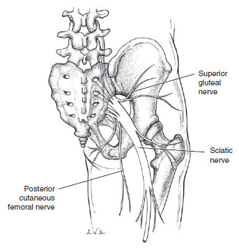
The large sciatic nerve (Figure 6-1) exits the pelvis through the greater sciatic foramen and enters the deep gluteal region immediately inferior to the piriformis muscle. The sciatic nerve is actually the combined common fibular nerve (lateral portion) and the tibial nerve (medial portion). The common fibular nerve innervates the lateral compartment muscles (superficial fibular branch) and the anterior compartment muscles (deep fibular branch) of the leg. The tibial portion innervates the muscles of posterior compartments of the thigh and leg (calf) and the sole of the foot. The sciatic nerve lies posterior to the hip joint.
|
Table 5-2 • INTRINSIC
MUSCLES OF THE SHOULDER
|
|
|
Muscles
|
Innervation
|
|
Flexion
|
Iliopsoas (psoas)
|
Anterior rami L1–L3 nerves
|
|
Iliopsoas (iliacus), rectus
femoris, pectineus, Sartorius
|
Femoral nerve
|
|
|
Adductor longus, brevis, and
magnus, gracilis
|
Obturator nerve
|
|
Tensor fascia lata
|
Superior gluteal nerve
|
|
Extension
|
Hamstrings: semitendinosus,
semimembranosus,
long head of biceps femoris,
adductor magnus (hamstring portion)
|
Tibial portion of sciatic nerve
|
|
Gluteus maximus
|
Inferior gluteal nerve
|
|
Abduction
|
Gluteus medius and minimus, tensor fascia
lata
|
Superior gluteal nerve
|
|
Adduction
|
Adductor longus, brevis and magnus,
gracilis, obturator externus
|
Obturator nerve
|
|
Pectineus
|
Femoral nerve
|
|
Medial rotation
|
Gluteus medius and minimus, tensor fascia
lata
|
Inferior gluteal nerve
|
|
Lateral rotation
|
Obturator internus, piriformis,
superior
and inferior gemelli, quadratus
femoris
|
Direct nerves to muscles arising
from L5, S1–S2
|
|
Obturator externus
|
Obturator nerve
|
|
Gluteus maximus
|
Inferior gluteal nerve
|

Figure 6-1. The sciatic nerve. (Reproduced, with permission, from Lindner HH. Clinical Anatomy. East Norwalk, CT: Appleton & Lange, 1989:591.)
COMPREHENSION QUESTIONS
6.1 In a patient who has a posterior dislocation of the hip, which of the following ligamentous structures would be torn?
A. Pubofemoral ligament
B. Iliofemoral ligament
C. Ischiofemoral ligament
D. Lacunar ligament
E. Sacrotuberous ligament
6.2 A 54-year-old man has just dislocated his right hip. The physician is concerned about the integrity of the joint’s blood supply. Which artery supplies most of the blood supply to the hip joint?
A. Lateral circumflex femoral
B. Medial circumflex femoral
C. Superficial circumflex iliac
D. Deep circumflex iliac
E. Perforating
6.3 A patient with a hip dislocation is also exhibiting weakness of extension of the thigh at the hip. This would indicate possible damage to which of the following?
A. Femoral nerve
B. Obturator nerve
C. Common fibular portion of the sciatic nerve
D. Tibial portion of the sciatic nerve
E. Saphenous nerve
Answers
6.1 C. Posterior dislocation of the hip would tear the ischiofemoral ligament, thus reinforcing the capsule of the hip posteriorly.
6.2 B. The chief blood supply to the hip is the medial circumflex femoral artery.
6.3 D. Most of the extensor muscles of the hip (the hamstrings) are innervated by the tibial portion of the sciatic nerve. The gluteus maximus muscle, innervated by the inferior gluteal nerve, could still weakly extend the thigh at the hip.
ANATOMY PEARLS
⯈ The bones forming the hip bone, ilium, ischium, and pubis converge to form the acetabulum.
⯈ The strongest ligament reinforcing the hip capsule is the iliofemoral or Y ligament.
⯈ The most important artery supplying the hip joint is the medial circumflex femoral artery.
References
REFERENCES
Gilroy AM, MacPherson BR, Ross LM. Atlas of Anatomy, 2nd ed. New York, NY: Thieme Medical
Publishers; 2012:386−389.
Moore KL, Dalley AF, Agur AMR. Clinically Oriented Anatomy, 7th ed. Baltimore, MD: Lippincott
Williams & Wilkins, 2014:626−634, 660−661.
Netter FH. Atlas of Human Anatomy, 6th ed. Philadelphia, PA: Saunders, 2014: plates 473−475, 490−491.


0 comments:
Post a Comment
Note: Only a member of this blog may post a comment.